
Stock image Rakusu
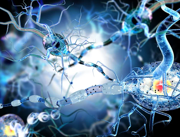
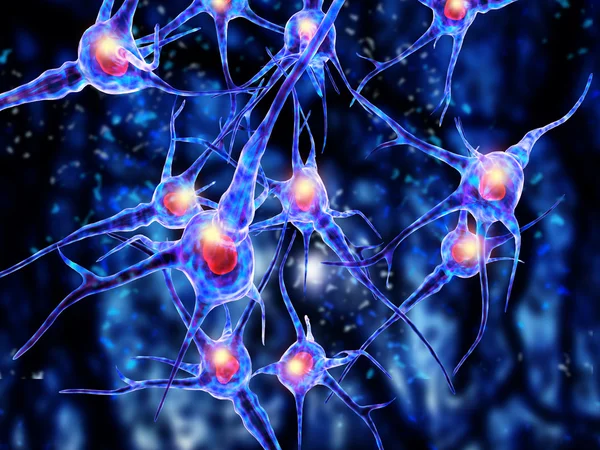
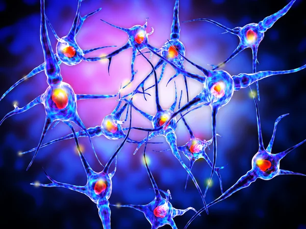
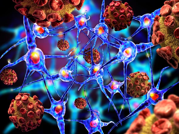
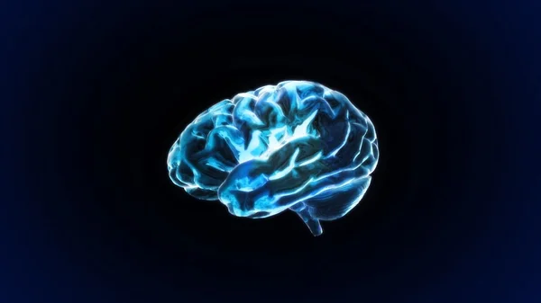
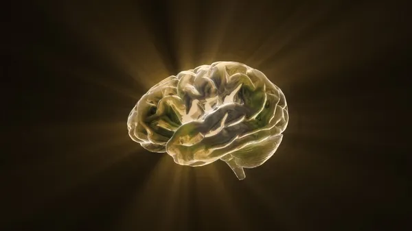
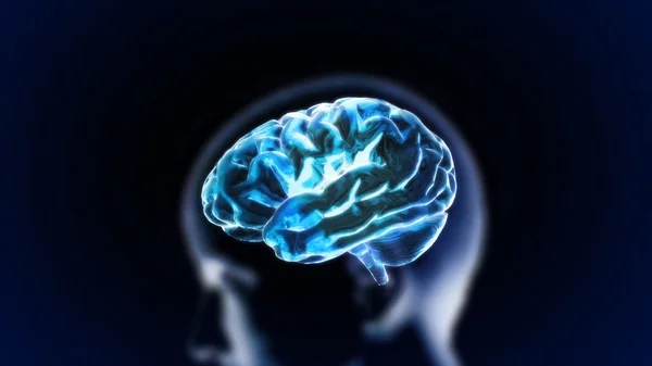
Nerve Cells, Concept For Neurological Diseases, Tumors And Brain Surgery.
Image, 9.49MB, 4712 × 3600 jpg
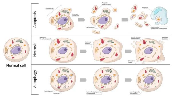
Three Basic Forms Of Cell Death: Apoptosis (chromosome Condensation, Nuclear Fragmentation), Autophagy (autophagosome Formation), Necrosis (membrane Rupture, Organelles Swelling).
Vector, 9.69MB, 7292 × 4167 eps


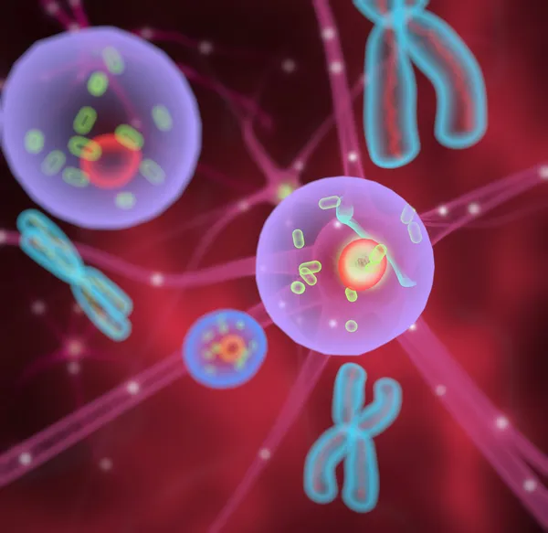
P53 Bound To DNA P53 Prevents Cancer Formation And Acts As A Guardian Of The Genome. Mutations In The P53 Gene Contribute To About Half Of The Cases Of Human Cancer. 3d Rendering. Illustration
Image, 4.94MB, 8000 × 6000 jpg
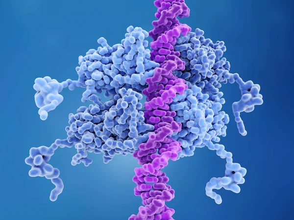
P53 Bound To DNA P53 Prevents Cancer Formation And Acts As A Guardian Of The Genome. Mutations In The P53 Gene Contribute To About Half Of The Cases Of Human Cancer. 3d Rendering. Illustration
Image, 4.36MB, 8000 × 6000 jpg
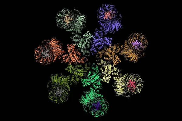
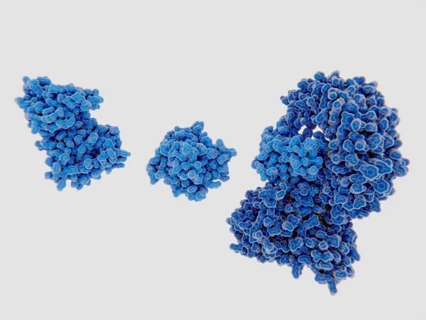
Structure Of The Apaf-1 Apoptosome With Cytochrome C Shown, 3D Cartoon Model, Black Background
Image, 3.17MB, 6000 × 4005 jpg

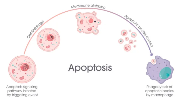
Apoptosis. Programmed Cell Death. Aging Process In Cells. Structural Changes Of Ageing And Senescent Cells From Normal Cell To Final Stage Of Formation Of Membrane Blebbing. Vector Illustration
Vector, 8.22MB, 5000 × 3352 eps
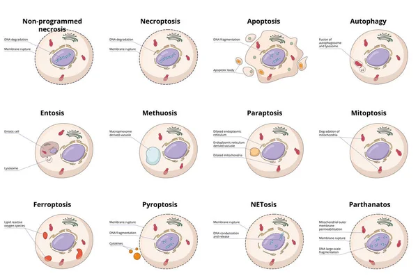
Cell Death Types: General Differences Between Cell Death Processes, Including Common Necrosis, Autophagy, Apoptosis And Specific Entosis, Paraptosis And Ferroptosis.
Vector, 10.32MB, 6250 × 4167 eps
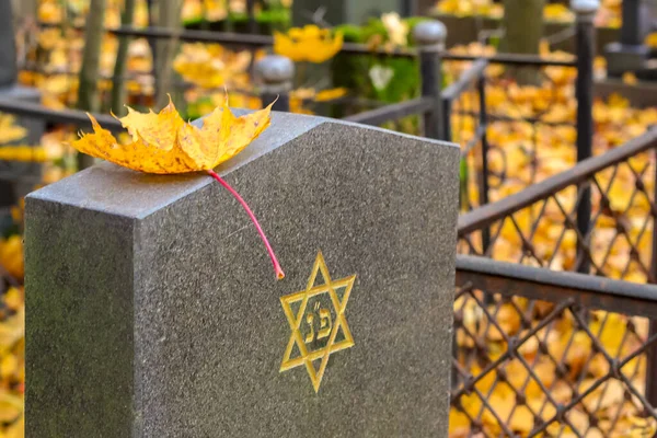
Autumn Jewish Cemetery. A Yellow Maple Leaf Is Lying On A Cemetery Plate With An Engraved Star Of David.
Image, 8.5MB, 4930 × 3287 jpg
The Mitotic Kinase NEK7 Binds To Inactive NRLP3 Leading To The Activation Of The Inflammasome
Image, 3.59MB, 8000 × 6000 jpg
Icons Set Premium Quality Of Biochemistry Research, Biology Laboratory Experiment. Modern Pictogram Collection Flat Design Style Symbol Collection. Isolated White Background.
Vector, 0.41MB, 5000 × 5600 eps
Icons Line Set Premium Quality Of Biochemistry Research, Biology Laboratory Experiment. Modern Pictogram Collection Flat Design Style Symbol . Isolated White Background
Vector, 0.54MB, 5000 × 5600 eps
FAST Stroke Symptoms Concept Icon. Typical Signs Of Stroke Idea Thin Line Illustration. Facial Drooping, Arm Weakness And Speech Difficulty. Vector Isolated Outline RGB Color Drawing. Editable Stroke
Vector, 0.31MB, 5000 × 5000 eps

T-cell Receptors Are Similar To One Arm Of An Antibody. Like Antibodies, They Are Composed Of Two Chains. The Binding Site Is At The Tip Of The Molecule,
Image, 2.5MB, 8000 × 6000 jpg
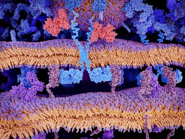
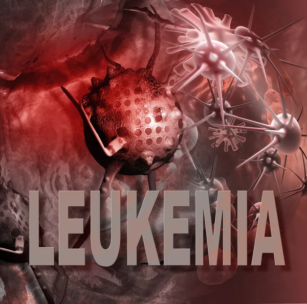
Engineered Receptors (light Blue) On The Surface Of A T-lymphocyte Bind Specifically To CD19-antigen Molecules (magenta) On A Leukemia Cell. This Activates A Signal Cascade In The T-cell Leading To The Segregation Of Vesicles That Contain Perforin An
Image, 11.44MB, 8000 × 6000 jpg

Chocolate Mousse Or Moldy Yogurt Has Expired. Woman's Hands Hold Plastic Cup With Chocolate Mousse Or Yogurt That Has Gone Bad And Moldy. Top View, White Background
Image, 4.76MB, 6000 × 4000 jpg
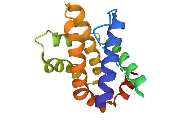
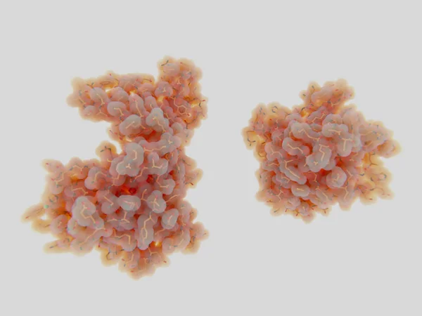
Structure Of Human Apoptosis Regulator Bcl-XL. 3D Cartoon Model, Sequence Id Color Scheme, PDB 1r2d, White Background
Image, 2.08MB, 6000 × 4000 jpg


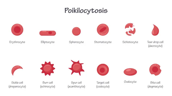
Poikilocytosis Morphology Of Erythrocytes Red Blood Cell RBC Educational Vector Illustration Graphic
Vector, 0.46MB, 8333 × 4688 ai


Page 1 >> Next