photo free
Home Images Vectors Videos

Stock image Ranvier page 2

stock vector Semicircular infographic with the possible symptoms of multiple sclerosis and its icons in white on orange tones.

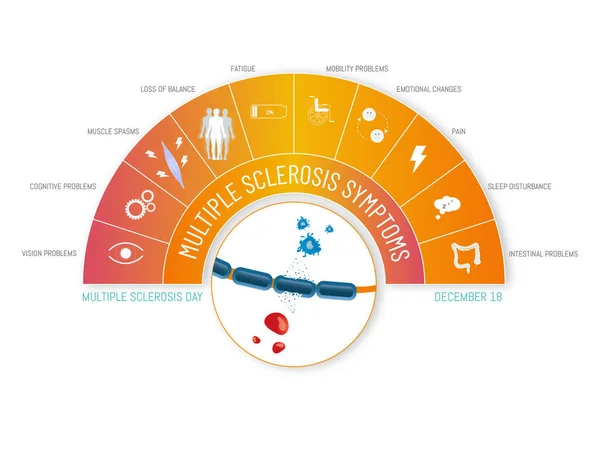
Semicircular Infographic With The Possible Symptoms Of Multiple Sclerosis And Its Icons In White On Orange Tones.

Vector, 8.47MB, 8333 × 6250 ai
stock image Transmission of nerve impulses with a zoom on the myelin sheath, Ranvier's node and the synapse.

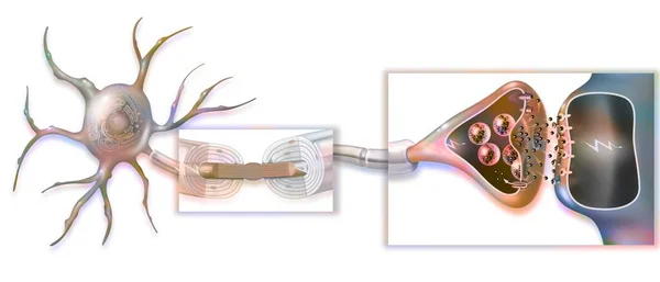
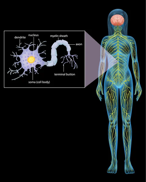
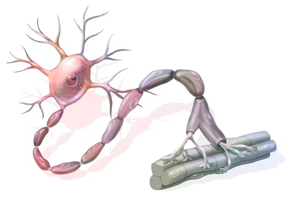
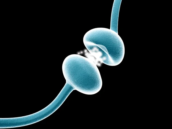
Transmission Of Nerve Impulses With A Zoom On The Myelin Sheath, Ranvier's Node And The Synapse.

Image, 1.39MB, 7048 × 3020 jpg
stock vector Multiple sclerosis. Autoimmune disease. Normal neuron and a neuron with a damaged myelin sheath. Nerve damage. Isometric Vector. Flat illustration

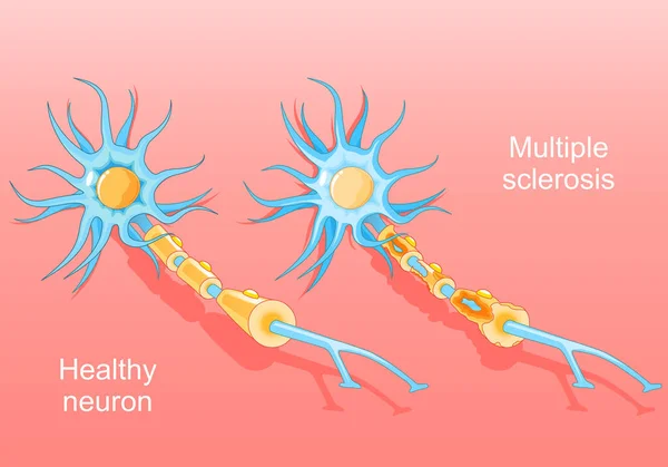
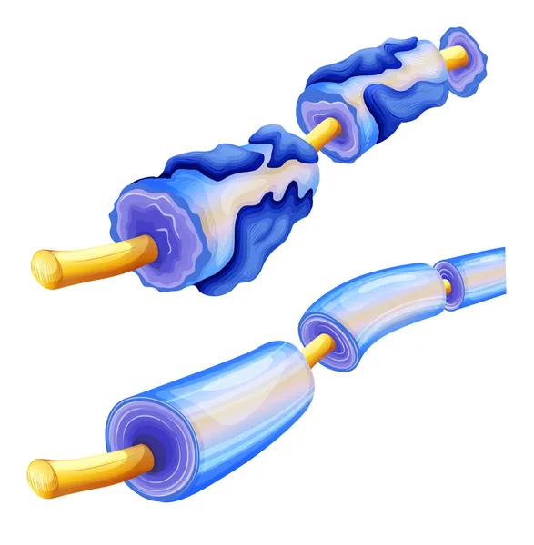
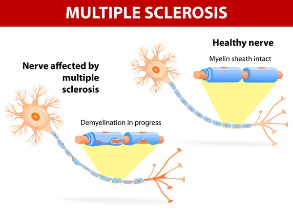
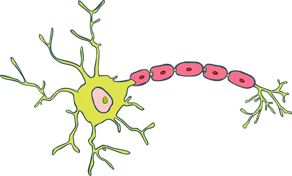
Multiple Sclerosis. Autoimmune Disease. Normal Neuron And A Neuron With A Damaged Myelin Sheath. Nerve Damage. Isometric Vector. Flat Illustration

Vector, 1.07MB, 5000 × 3498 eps
stock vector Anatomy of a typical human neuron  Vector

Anatomy Of A Typical Human Neuron Vector

Vector, 3.46MB, 8190 × 8190 eps
stock vector Human Neuron

Human Neuron

Vector, 10.02MB, 5000 × 5530 eps
stock vector Female Neuron

Female Neuron

Vector, 7.75MB, 4000 × 4992 eps
stock vector Vector neuron scheme

Vector Neuron Scheme

Vector, 5.79MB, 4244 × 6001 eps
stock image White matter: neurons with myelinated axons, oligodendrocytes forming the myelin sheaths, fibrous astrocytes and microglia cells

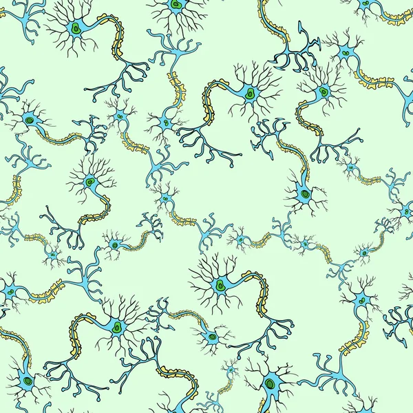
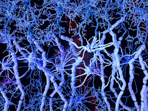
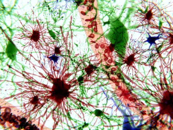
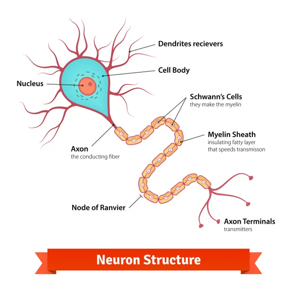
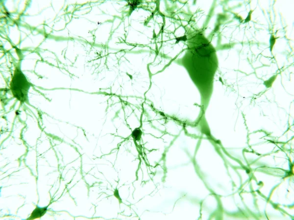
White Matter: Neurons With Myelinated Axons, Oligodendrocytes Forming The Myelin Sheaths, Fibrous Astrocytes And Microglia Cells

Image, 12.77MB, 8000 × 6000 jpg
stock image Oligodendrocytes form insulating myelin sheaths around neuron axons in the central nervous system. One oligodendrocyte can extend its processes to 50 axons. Myelin increases the impulse speed and decreases the capacitance of the axon membrane.

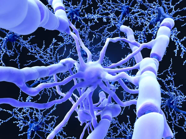
Oligodendrocytes Form Insulating Myelin Sheaths Around Neuron Axons In The Central Nervous System. One Oligodendrocyte Can Extend Its Processes To 50 Axons. Myelin Increases The Impulse Speed And Decreases The Capacitance Of The Axon Membrane.

Image, 8.09MB, 8000 × 6000 jpg
stock image Brain cells: astrocytes (red), pyramidal neurons (red), microglia cells (blue)

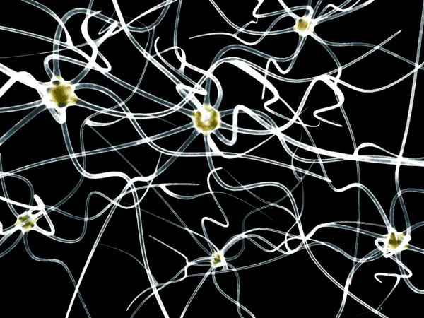
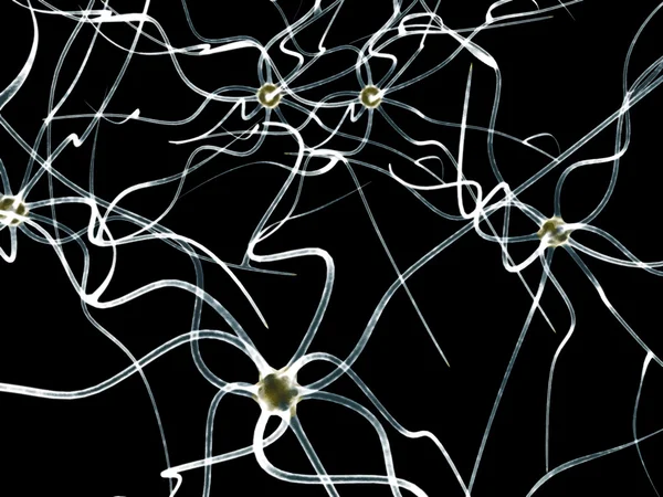
Brain Cells: Astrocytes (red), Pyramidal Neurons (red), Microglia Cells (blue)

Image, 8.6MB, 8000 × 6000 jpg
stock vector Neuron cell

Neuron Cell

Vector, 0.34MB, 2457 × 3227 eps
stock vector Digital illustration of neuron Vector

Digital Illustration Of Neuron Vector

Vector, 13.21MB, 8100 × 6075 eps
stock vector Myelin sheath layer for axon nerve with detailed structure outline diagram. Labeled educational anatomy scheme with microbiological medical nervous system part vector illustration. Neuron insulation.

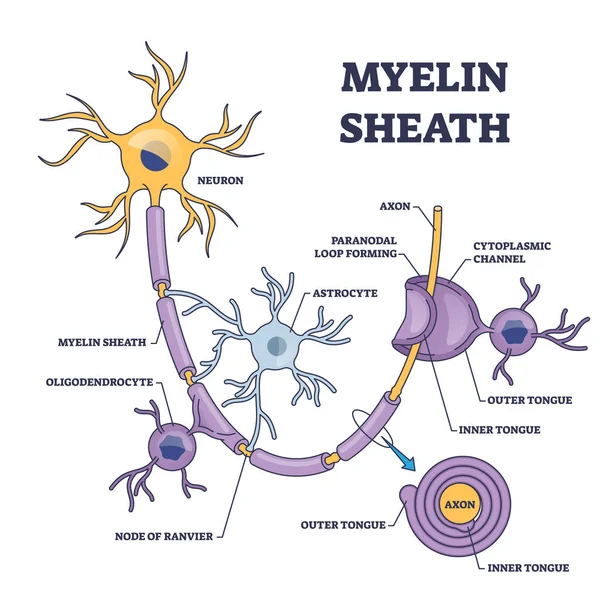
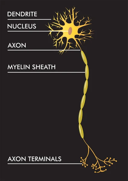
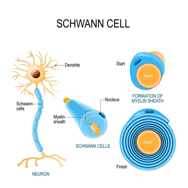
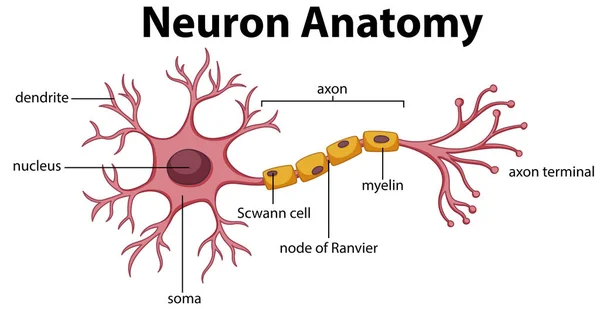
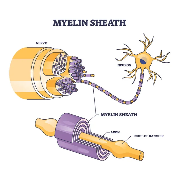
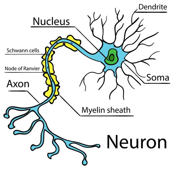
Myelin Sheath Layer For Axon Nerve With Detailed Structure Outline Diagram. Labeled Educational Anatomy Scheme With Microbiological Medical Nervous System Part Vector Illustration. Neuron Insulation.

Vector, 6.46MB, 4200 × 4200 eps
stock image Motor neuron: neuron in contact with muscle fibers.

Motor Neuron: Neuron In Contact With Muscle Fibers.

Image, 0.81MB, 4394 × 3020 jpg
stock vector Human Neuron Anatomy

Human Neuron Anatomy

Vector, 1.93MB, 7000 × 3478 eps
stock image Book with diagnosis   autoimmune diseases.

Book With Diagnosis Autoimmune Diseases.

Image, 8.96MB, 5500 × 3667 jpg
stock vector Infographic of a healthy neuron and one with damage from attack by myelin-destroying lymphocytes and macrophages in multiple sclerosis disease.

Infographic Of A Healthy Neuron And One With Damage From Attack By Myelin-destroying Lymphocytes And Macrophages In Multiple Sclerosis Disease.

Vector, 1.73MB, 8333 × 6250 ai
stock vector Anatomy of a typical human neuron Vector 1

Anatomy Of A Typical Human Neuron Vector 1

Vector, 14.8MB, 8000 × 6080 eps
stock vector Brain neuron cell diagram

Brain Neuron Cell Diagram

Vector, 2.94MB, 4000 × 4000 eps
stock vector Digital illustration of neuron Vector

Digital Illustration Of Neuron Vector

Vector, 11.66MB, 8100 × 6075 eps
stock vector The structure of the pyromidal neuron. Nerve cell. Infographics. Vector illustration on isolated background

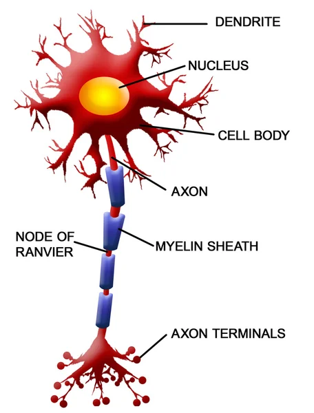
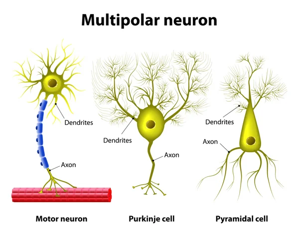
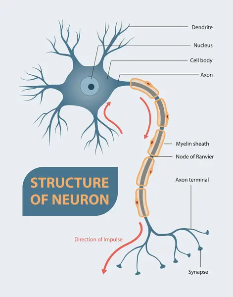
The Structure Of The Pyromidal Neuron. Nerve Cell. Infographics. Vector Illustration On Isolated Background

Vector, 1.37MB, 5000 × 5000 eps
stock vector Betz cell. Neuron. Nerve cell. Infographics. Vector illustration on isolated background.

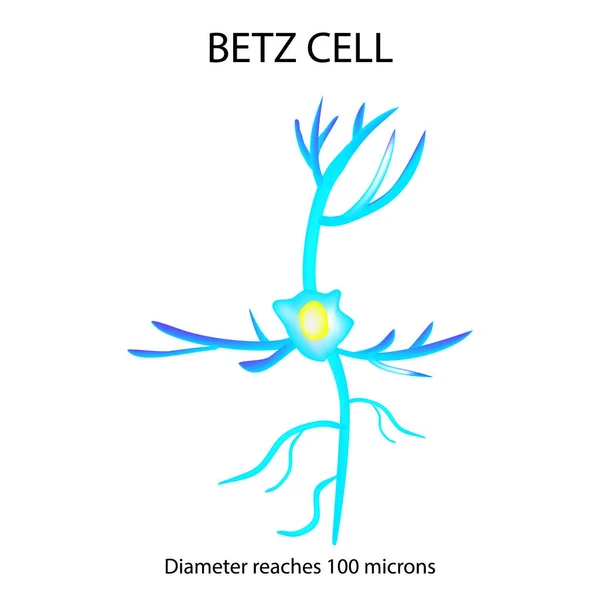
Betz Cell. Neuron. Nerve Cell. Infographics. Vector Illustration On Isolated Background.

Vector, 1.16MB, 5000 × 5000 eps
stock image Autoimmune Attack on Myelin Sheath 3D rendering

Autoimmune Attack On Myelin Sheath 3D Rendering

Image, 10.21MB, 7258 × 3920 jpg
stock vector Types Of a multipolar Neurons

Types Of A Multipolar Neurons

Vector, 3.24MB, 5346 × 4209 eps
stock vector Anatomy of a typical human neuron Vector3

Anatomy Of A Typical Human Neuron Vector3

Vector, 9.65MB, 8100 × 6075 eps
stock vector Schwann cells. Structure of neurolemmocytes.

Schwann Cells. Structure Of Neurolemmocytes.

Vector, 1.79MB, 5327 × 5327 eps
stock vector Diagram of Neuron Anatomy  illustration

Diagram Of Neuron Anatomy Illustration

Vector, 1.06MB, 6532 × 3369 eps
stock image Nerve impulse: central nervous system neural network with zoom on Ranvier's node.

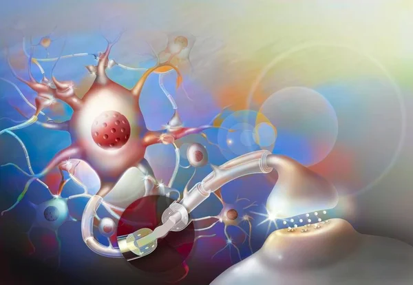
Nerve Impulse: Central Nervous System Neural Network With Zoom On Ranvier's Node.

Image, 1.76MB, 5079 × 3514 jpg
stock image Neuron anatomy isolated in black background.3d illustration

Neuron Anatomy Isolated In Black Background.3d Illustration

Image, 1.56MB, 3840 × 2160 jpg
stock vector Neuron cell

Neuron Cell

Vector, 0.29MB, 2457 × 3227 eps
stock vector Anatomy of a typical human neuron Vector 2

Anatomy Of A Typical Human Neuron Vector 2

Vector, 6.15MB, 8191 × 6143 eps
stock vector Myelin sheath as insulation layer for brain or spinal nerve outline diagram. Labeled educational anatomical scheme with physiological neuron, axon and node of ranvier location vector illustration.

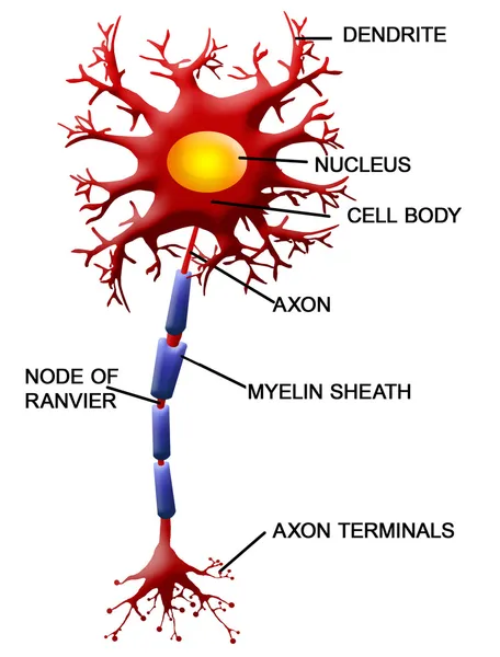
Myelin Sheath As Insulation Layer For Brain Or Spinal Nerve Outline Diagram. Labeled Educational Anatomical Scheme With Physiological Neuron, Axon And Node Of Ranvier Location Vector Illustration.

Vector, 6.22MB, 4200 × 4200 eps
stock image Postsynaptic neuron receptors (dendrite) receive emitted neurotransmitters from presynaptic neuron (axon) - isometric view 3d illustration

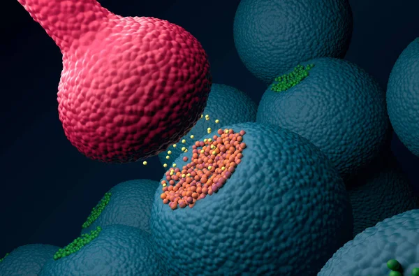
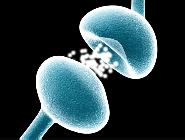
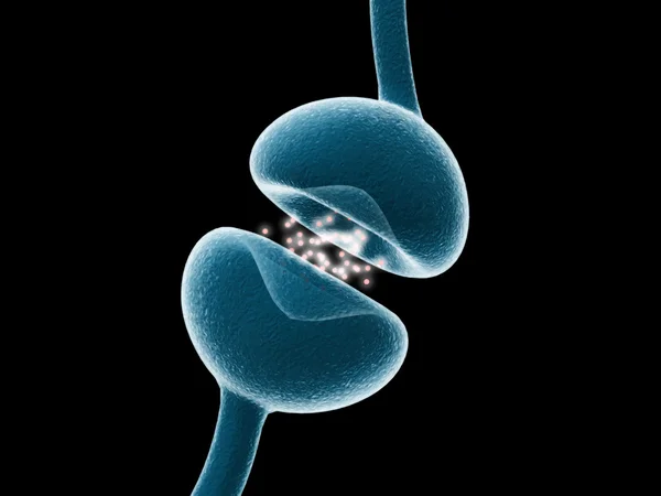
Postsynaptic Neuron Receptors (dendrite) Receive Emitted Neurotransmitters From Presynaptic Neuron (axon) - Isometric View 3d Illustration

Image, 8.03MB, 10000 × 6600 jpg
stock vector Myelin deterioration

Myelin Deterioration

Vector, 7.37MB, 4892 × 5000 eps
stock vector Nerve affected by multiple sclerosis

Nerve Affected By Multiple Sclerosis

Vector, 6.92MB, 4976 × 3638 eps
stock image Multiple Sclerosis Concept

Multiple Sclerosis Concept

Image, 5.28MB, 4096 × 2616 jpg
stock vector Anatomy of a typical human neuron

Anatomy Of A Typical Human Neuron

Vector, 4.14MB, 3306 × 2000 eps
stock vector Structure of neuron Illustration

Structure Of Neuron Illustration

Vector, 5.92MB, 5758 × 7336 eps
stock vector Digital illustration of neuron Vector

Digital Illustration Of Neuron Vector

Vector, 11.29MB, 8100 × 6075 eps
stock image Human pyramidal neurons. Illustration

Human Pyramidal Neurons. Illustration

Image, 2.6MB, 8000 × 6000 jpg
stock vector Neuron icon. Vector

Neuron Icon. Vector

Vector, 0.74MB, 5000 × 5000 eps
stock image Multiple Sclerosis Concept

Multiple Sclerosis Concept

Image, 4.48MB, 4096 × 2616 jpg
stock vector Anatomy of a typical human neuron Vector 2

Anatomy Of A Typical Human Neuron Vector 2

Vector, 0.94MB, 8190 × 8190 eps
Previous << Page 2 >> Next