photo free
Home Images Vectors Videos

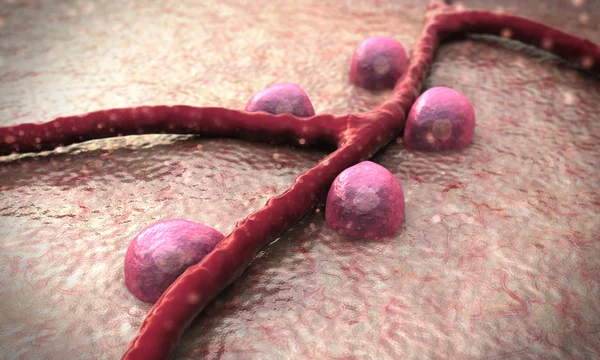
Stock image Receptor Protein

stock image The amyloid precursor protein. When cleaved, the membrane domain is involved in the Alzheimer disease building amyloid plaques. 3d rendering. Illustration

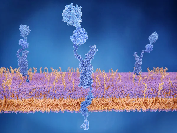
The Amyloid Precursor Protein. When Cleaved, The Membrane Domain Is Involved In The Alzheimer Disease Building Amyloid Plaques. 3d Rendering. Illustration

Image, 3.29MB, 8000 × 6000 jpg
stock image The angiotensin converting enzyme 2 (ACE2, blue) in complex with the amino acid transporter BOAT 1 (violet). ACE2 is involved in control of blood pressure and is the target for the SARS-CoV-2 virus to infect human cells.

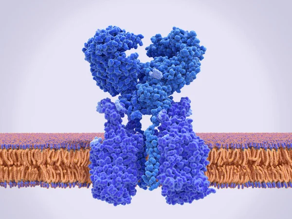
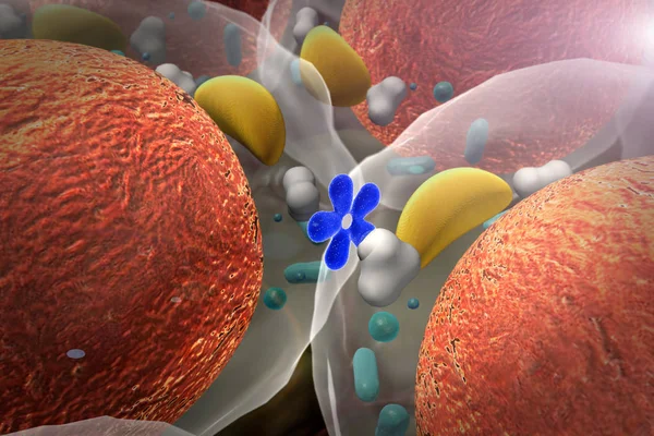
The Angiotensin Converting Enzyme 2 (ACE2, Blue) In Complex With The Amino Acid Transporter BOAT 1 (violet). ACE2 Is Involved In Control Of Blood Pressure And Is The Target For The SARS-CoV-2 Virus To Infect Human Cells.

Image, 4.5MB, 8000 × 6000 jpg
stock image The amyloid precursor protein (APP) is a complex protein with many functions. It is found on the surface of cells throughout the body. The intact protein binds to many stuctural proteins outside cells, such as heparin and laminin and sends signals th

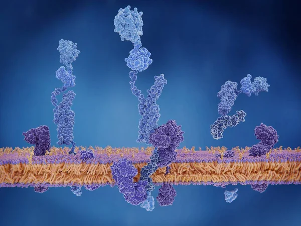
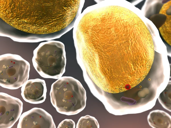
The Amyloid Precursor Protein (APP) Is A Complex Protein With Many Functions. It Is Found On The Surface Of Cells Throughout The Body. The Intact Protein Binds To Many Stuctural Proteins Outside Cells, Such As Heparin And Laminin And Sends Signals Th

Image, 6.49MB, 8000 × 6000 jpg
stock vector Hormones, receptors and target Cells

Hormones, Receptors And Target Cells

Vector, 10.44MB, 4896 × 4385 eps
stock vector Animal cell with nucleus, cytoplasm, DNA, enzime, protein kinase

Animal Cell With Nucleus, Cytoplasm, DNA, Enzime, Protein Kinase

Vector, 1.14MB, 5421 × 5421 eps
stock image Receptors on the cells surface

Receptors On The Cells Surface

Image, 7.35MB, 5000 × 3750 jpg
stock vector Steroid hormones response. Vector

Steroid Hormones Response. Vector

Vector, 6.57MB, 5086 × 4211 eps
stock image 3D image of tyrosine skeletal formula - molecular chemical structure of 4-hydroxyphenylalanine isolated on white background

3D Image Of Tyrosine Skeletal Formula - Molecular Chemical Structure Of 4-hydroxyphenylalanine Isolated On White Background

Image, 2.01MB, 5500 × 3630 jpg
stock image 3D image of Neuropeptide Y skeletal formula - molecular chemical structure of vasoconstrictor isolated on white background

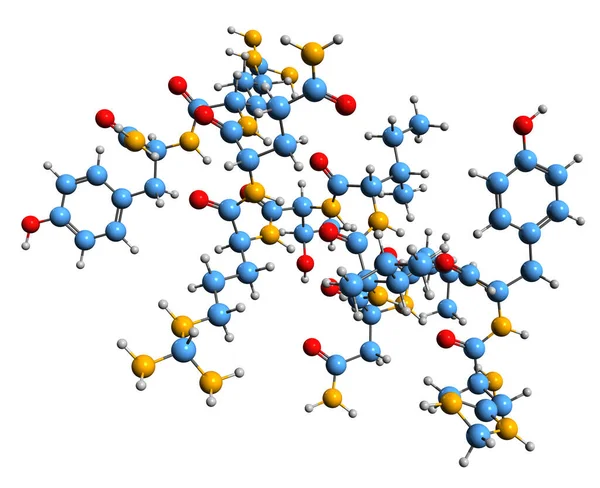
3D Image Of Neuropeptide Y Skeletal Formula - Molecular Chemical Structure Of Vasoconstrictor Isolated On White Background

Image, 6.87MB, 8179 × 6660 jpg
stock image 3D image of triiodothyronine skeletal formula - molecular chemical structure of thyroid hormone T3 isolated on white background

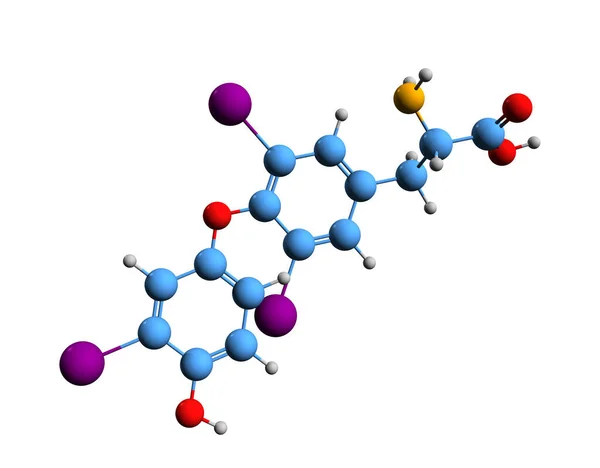
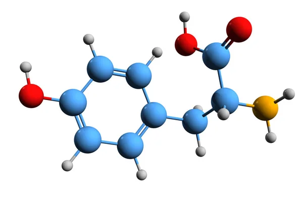
3D Image Of Triiodothyronine Skeletal Formula - Molecular Chemical Structure Of Thyroid Hormone T3 Isolated On White Background

Image, 2.21MB, 5500 × 4296 jpg
stock vector Cytokine receptor. signal transduction between cells.

Cytokine Receptor. Signal Transduction Between Cells.

Vector, 7.29MB, 4602 × 4601 eps
stock vector Anatomy of the Lysosome.

Anatomy Of The Lysosome.

Vector, 3.83MB, 3944 × 3944 eps
stock vector Ovarian cyst, follicle and Corpus luteum.

Ovarian Cyst, Follicle And Corpus Luteum.

Vector, 5.57MB, 7633 × 7221 eps
stock vector Cytotoxic cells. Cytokines. Cell immunity. Infographics. Vector illustration

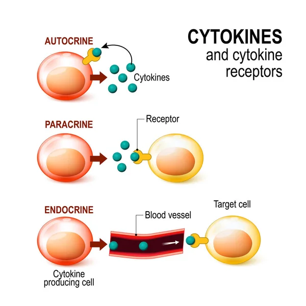
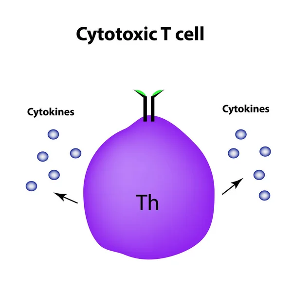
Cytotoxic Cells. Cytokines. Cell Immunity. Infographics. Vector Illustration

Vector, 1.39MB, 5000 × 5000 eps
stock image cholesterol in a cells

Cholesterol In A Cells

Image, 19.86MB, 8000 × 6000 jpg
stock image Fat cell, cholesterol

Fat Cell, Cholesterol

Image, 28.05MB, 8000 × 6000 jpg
stock image  cholesterol in a cells

Cholesterol In A Cells

Image, 8.96MB, 6000 × 4000 jpg
stock vector Insulin in healthy body brings glucose into the blood stream for

Insulin In Healthy Body Brings Glucose Into The Blood Stream For

Vector, 17.58MB, 2808 × 5000 eps
stock vector endocytosis. phagocytosis is cell eating, pinocytosis is a cell drinking, receptor-mediated endocytosis - when cells absorb metabolites, hormones, proteins and viruses by receptors on the surface of the cell.

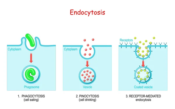
Endocytosis. Phagocytosis Is Cell Eating, Pinocytosis Is A Cell Drinking, Receptor-mediated Endocytosis - When Cells Absorb Metabolites, Hormones, Proteins And Viruses By Receptors On The Surface Of The Cell.

Vector, 1.03MB, 5357 × 3333 eps
stock image Fat cells

Fat Cells

Image, 13.11MB, 8000 × 6000 jpg
stock image Cholesterol, cell

Cholesterol, Cell

Image, 22.29MB, 8000 × 6000 jpg
stock image Fat cells, cell

Fat Cells, Cell

Image, 29.34MB, 8000 × 6000 jpg
stock vector Low insulin levels.

Low Insulin Levels.

Vector, 19.3MB, 6000 × 6000 eps
stock vector GABA receptor. ligand-gated ion channel, metabotropic receptors. Neurotransmitter in the central nervous system.

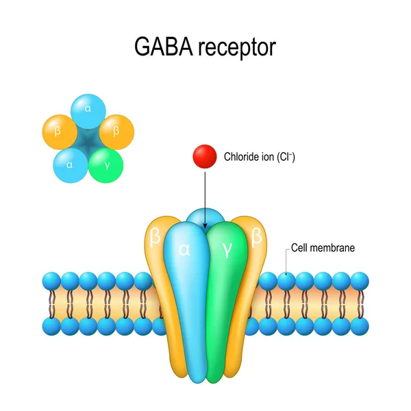
GABA Receptor. Ligand-gated Ion Channel, Metabotropic Receptors. Neurotransmitter In The Central Nervous System.

Vector, 12.32MB, 4444 × 4444 eps
stock image  cholesterol in a cells

Cholesterol In A Cells

Image, 10.37MB, 7000 × 5250 jpg
stock vector Membrane proteins labeled vector illustration. Detailed structure scheme.

Membrane Proteins Labeled Vector Illustration. Detailed Structure Scheme.

Vector, 8.91MB, 4100 × 3972 eps
stock vector The life cycle of HIV. Infographics. World AIDS Day. Vector illustration

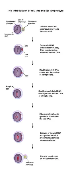
The Life Cycle Of HIV. Infographics. World AIDS Day. Vector Illustration

Vector, 9.28MB, 3276 × 8190 eps
stock image Turquiose neurone closeup

Turquiose Neurone Closeup

Image, 4.85MB, 4500 × 3000 jpg
stock vector Endocytosis. Lysosome digesting food. Part of cell (plasma membrane, cytoplasm and lysosome), with food vacuole. Lysosome fusing with the food vacuole. Vector illustration

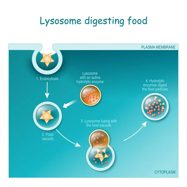
Endocytosis. Lysosome Digesting Food. Part Of Cell (plasma Membrane, Cytoplasm And Lysosome), With Food Vacuole. Lysosome Fusing With The Food Vacuole. Vector Illustration

Vector, 9.52MB, 4444 × 4444 eps
stock image Cholesterol, cell structure

Cholesterol, Cell Structure

Image, 18.04MB, 8000 × 6000 jpg
stock vector Cell membrane. GABA receptor. Top view of ion channel

Cell Membrane. GABA Receptor. Top View Of Ion Channel

Vector, 3.02MB, 5168 × 5168 eps
stock vector Lysosome. appearance, exterior and interior view. Cross section and Anatomy of the Lysosome: Hydrolytic enzymes, Membrane and transport proteins. Vector illustration

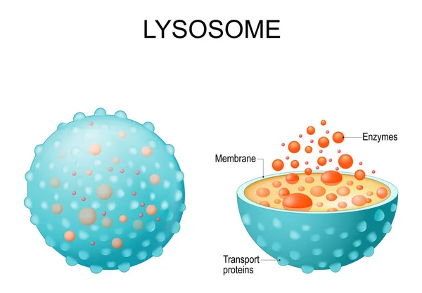
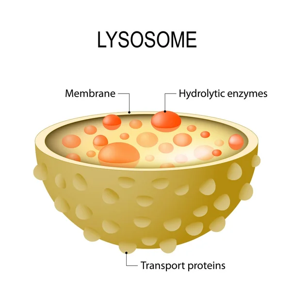
Lysosome. Appearance, Exterior And Interior View. Cross Section And Anatomy Of The Lysosome: Hydrolytic Enzymes, Membrane And Transport Proteins. Vector Illustration

Vector, 5.9MB, 5000 × 3482 eps
stock vector Bone Biology. Role of RANK, RANKL, and OPG. bone remodeling. Bone is broken down by osteoclasts, and rebuilt by osteoblasts. Receptor activator of RANKL is the mediator of bone resorption. Osteoprotegerin (OPG). Paracrine and endocrine actions of bon

Bone Biology. Role Of RANK, RANKL, And OPG. Bone Remodeling. Bone Is Broken Down By Osteoclasts, And Rebuilt By Osteoblasts. Receptor Activator Of RANKL Is The Mediator Of Bone Resorption. Osteoprotegerin (OPG). Paracrine And Endocrine Actions Of Bon

Vector, 13.41MB, 5742 × 3333 eps
stock vector Hormones, Receptors and Target Cells. each type of hormone is designed only certain cells. These cells will have receptors on them that are specific for a certain hormone. Vector illustration for medical, biological, and educational use

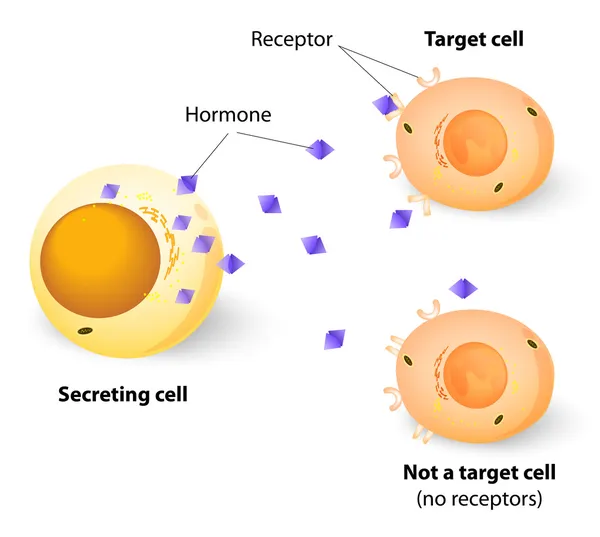
Hormones, Receptors And Target Cells. Each Type Of Hormone Is Designed Only Certain Cells. These Cells Will Have Receptors On Them That Are Specific For A Certain Hormone. Vector Illustration For Medical, Biological, And Educational Use

Vector, 2.77MB, 5013 × 5012 eps
stock image Cells, Macrophage

Cells, Macrophage

Image, 14.41MB, 8000 × 6000 jpg
stock image Fat cell, cells

Fat Cell, Cells

Image, 33.17MB, 8000 × 6000 jpg
stock image insulin enters the cell

Insulin Enters The Cell

Image, 6.81MB, 6000 × 4000 jpg
stock image Molecular model of Pembrolizumab, a humanized antibody used in immunotherapy of cancer, 3D illustration. It targets the PD-1 receptor of lymphocytes

Molecular Model Of Pembrolizumab, A Humanized Antibody Used In Immunotherapy Of Cancer, 3D Illustration. It Targets The PD-1 Receptor Of Lymphocytes

Image, 5.69MB, 6405 × 4269 jpg
stock image field of fat cells

Field Of Fat Cells

Image, 16.53MB, 8000 × 6000 jpg
stock image Ion Channels of a Cell

Ion Channels Of A Cell

Image, 4.47MB, 4300 × 3600 jpg
stock image gland

Gland

Image, 11.63MB, 6000 × 4000 jpg
stock image Neuron

Neuron

Image, 7.86MB, 8000 × 5000 jpg
stock image Metabolic Diseases concept

Metabolic Diseases Concept

Image, 2.43MB, 4500 × 3000 jpg
stock image hormone enters the cell

Hormone Enters The Cell

Image, 7.04MB, 6000 × 4000 jpg
stock image Closeup of purple neuron

Closeup Of Purple Neuron

Image, 5.04MB, 4500 × 3000 jpg
stock image Field of fat cells

Field Of Fat Cells

Image, 8.92MB, 4587 × 5000 jpg
stock image Cells, Macrophage

Cells, Macrophage

Image, 10.41MB, 8000 × 5000 jpg
stock image Beta cells, insulin

Beta Cells, Insulin

Image, 4.86MB, 5000 × 3000 jpg
stock vector Anatomy of the Lysosome: Hydrolytic enzymes, Membrane and transport  proteins. This organelle use the enzymes to break down and digest food particles, engulfed viruses or bacteria in the cell. Vector diagram for medical use

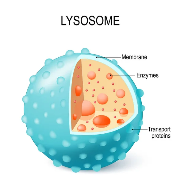
Anatomy Of The Lysosome: Hydrolytic Enzymes, Membrane And Transport Proteins. This Organelle Use The Enzymes To Break Down And Digest Food Particles, Engulfed Viruses Or Bacteria In The Cell. Vector Diagram For Medical Use

Vector, 6.8MB, 4230 × 4230 eps
stock vector Endocytosis process with closeup cell side view in anatomical outline diagram

Endocytosis Process With Closeup Cell Side View In Anatomical Outline Diagram

Vector, 5.64MB, 5000 × 3333 eps
Page 1 >> Next