
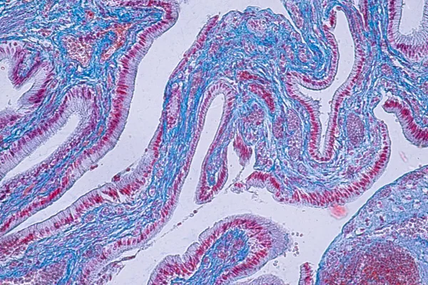
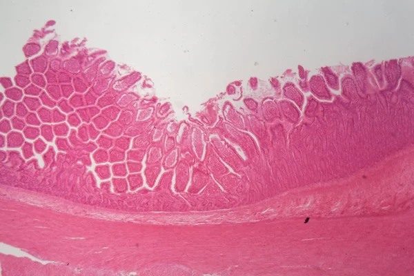
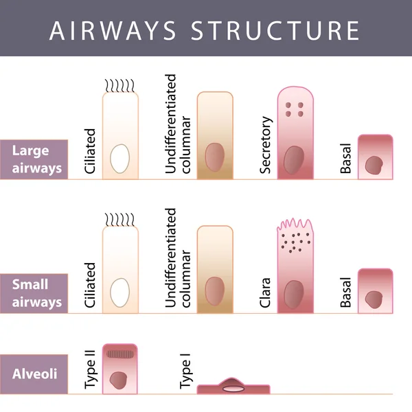
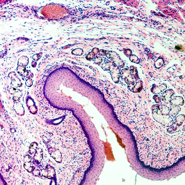
Stock image Respiratory Epithelium

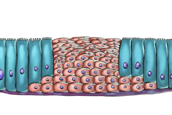
Pseudostratified Epithelium Is A Type Of Epithelium That, Though Comprising Only A Single Layer Of Cells.
Image, 11.24MB, 5840 × 3893 jpg


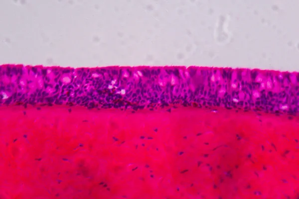

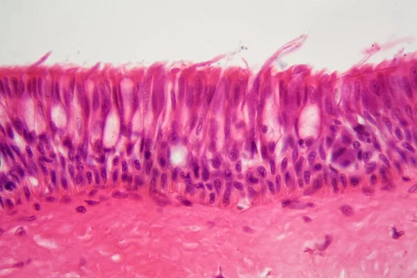
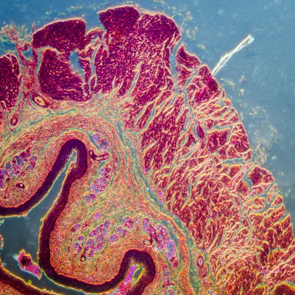
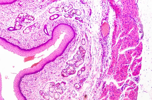
Cross Section Of Ciliated Epithelium Under The Microscope For Education Histology. Human Tissue.
Image, 14.75MB, 5168 × 3448 jpg
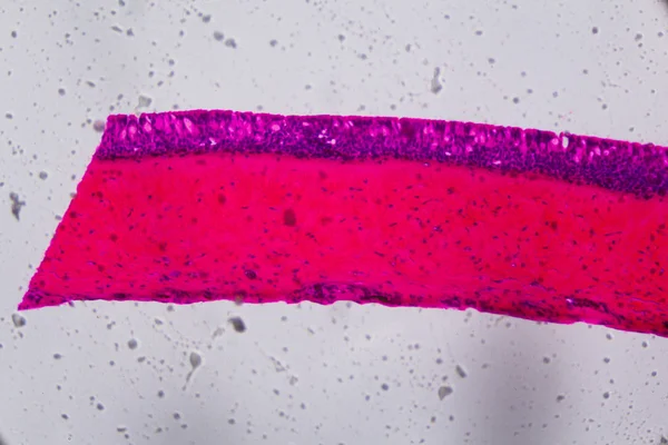


Ciliated Columnar Epithelium. Epithelial Cells Forms The Lining Of The Stomach And Intestines, Duodenum, Fallopian Tubes, Uterus, Central Canal Of The Spinal Cord, Nose, Ears And The Taste Buds.
Vector, 0.98MB, 4444 × 4444 eps


Structure Goblet Cells Of The Intestine. Infographics. Vector Illustration On Isolated Background
Vector, 3.04MB, 5000 × 5000 eps

Tonsil Stones Vector Illustration. Labeled Medical Tonsillolith Symptoms.
Vector, 6.56MB, 4500 × 3780 eps
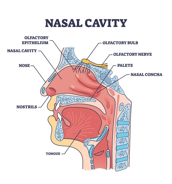
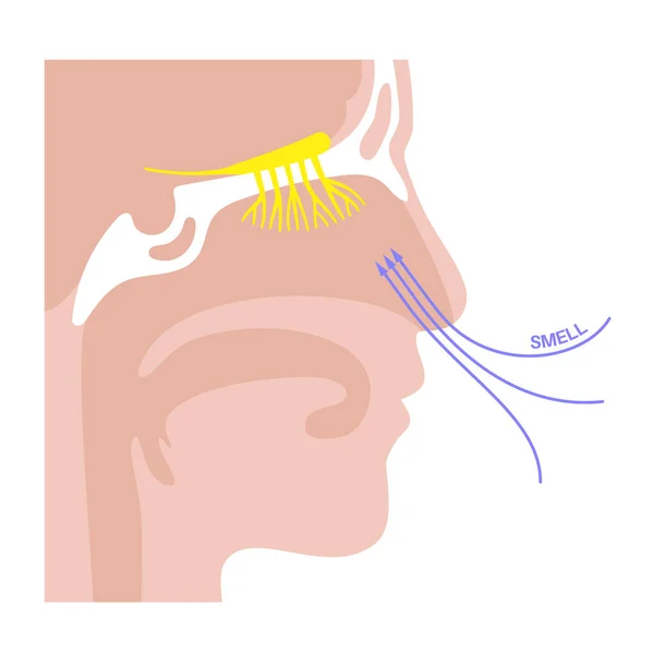
Nasal Cavity Anatomy With Medical Nose Parts Description Outline Diagram. Labeled Educational Cross Section Scheme With Mouth And Oral Structure Vector Illustration. Nostrils And Olfactory Location.
Vector, 5.92MB, 4000 × 4267 eps

Innate Immune System. Anatomical Barriers. Man Silhouette With Internal Organs. Blood Brain Barrier Protects The Nervous System From Pathogens. Shedding O
Vector, 12.03MB, 4444 × 4444 eps
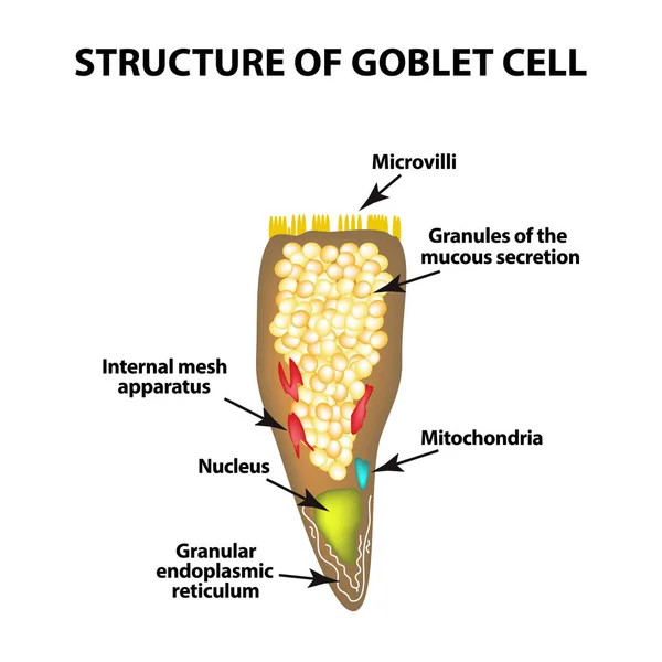
Structure Goblet Cells Of The Intestine. Infographics. Vector Illustration On Isolated Background
Vector, 3.04MB, 5000 × 5000 eps


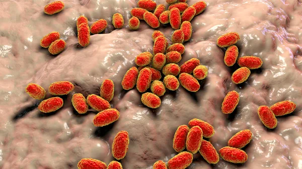
Whooping Cough Bacteria Bordetella Pertussis, 3D Illustration. Gram-negative Coccobacilli Bacteria Which Cause Children Infection Whooping Cough
Image, 14.57MB, 7200 × 4050 jpg


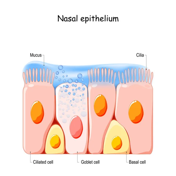
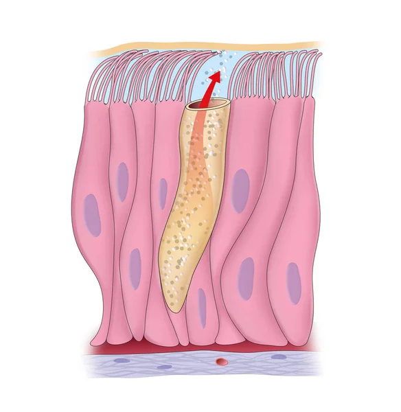
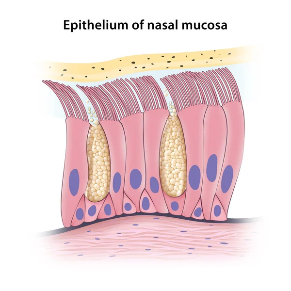
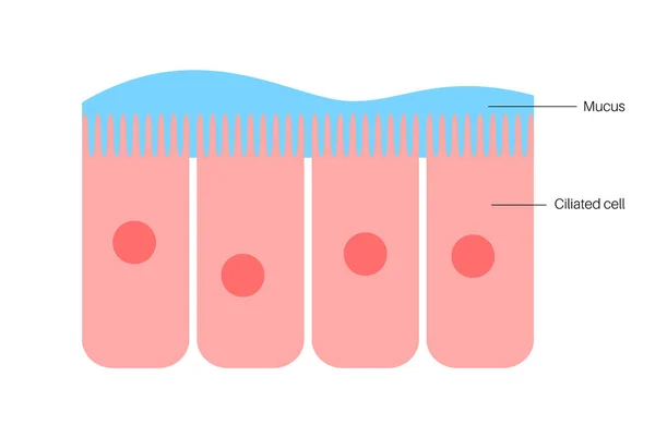
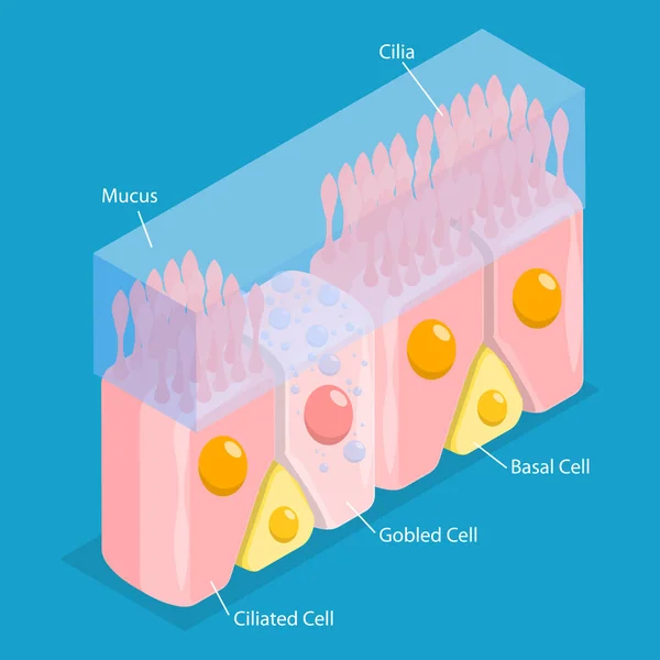
Nasal Mucosa Cells. Nasal Secretions. Ciliated, Basal And Goblet Cells. Olfactory Epithelium. Cells Act As A Low Resistance Filter. Vector Illustration
Vector, 11.58MB, 4444 × 4444 eps

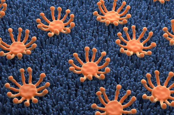
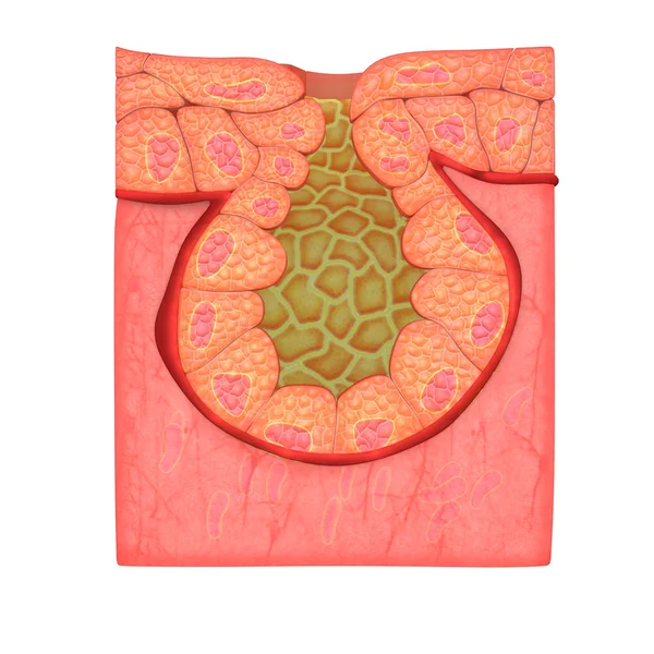
Smell (olfactory) Receptor Field In Nasal Lining - Isometric View 3d Illustration
Image, 12.55MB, 10000 × 6600 jpg


Mucociliary Transport System Black And White Simple Vector Icon, Medical Illustration
Vector, 0.52MB, 5000 × 5000 eps

Whooping Cough Bacteria Bordetella Pertussis, 3D Illustration. Gram-negative Coccobacilli Bacteria Which Cause Children Infection Whooping Cough
Image, 25.03MB, 7200 × 4050 jpg


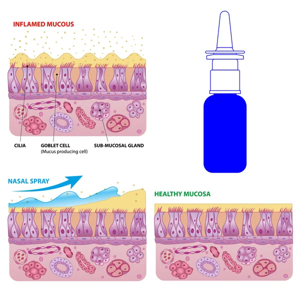
3D Isometric Flat Vector Conceptual Illustration Of Nasal Mucosa Cells, Medical Educational Diagram
Vector, 1.89MB, 5000 × 5000 eps
Page 1 >> Next