
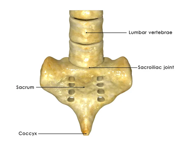
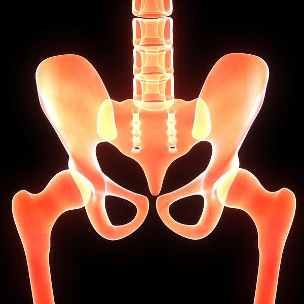
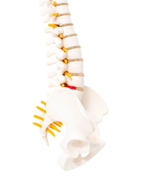
Stock image Sacroiliac Joint
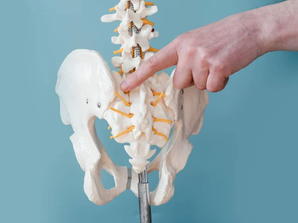
Close Up Of Male Doctors Hand Pointing At Sacroiliac Joint On Skeleton Spine Model
Image, 7.1MB, 5188 × 3896 jpg
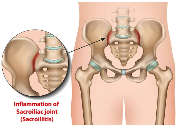
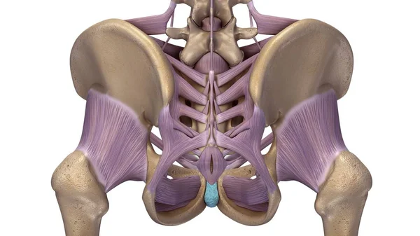
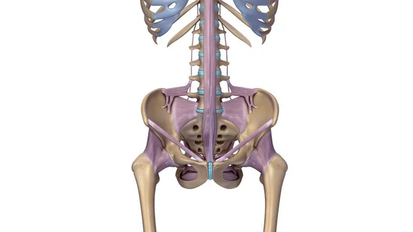
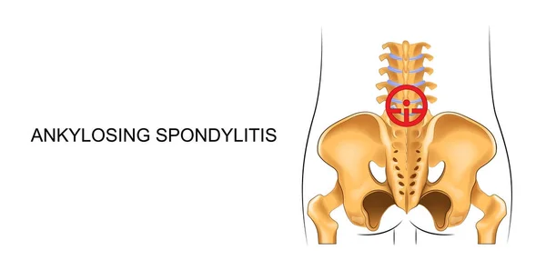
Sacroiliac Joint Inflammation 3d Medical Vector Illustration Sacroiliitis
Vector, 6.7MB, 7000 × 5000 eps
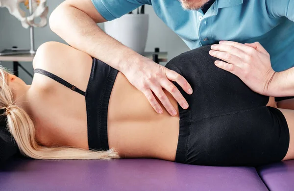
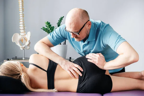
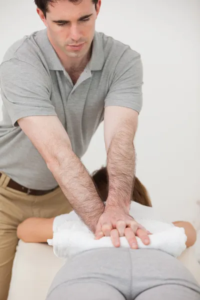
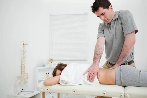
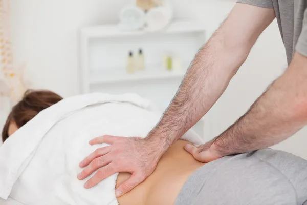
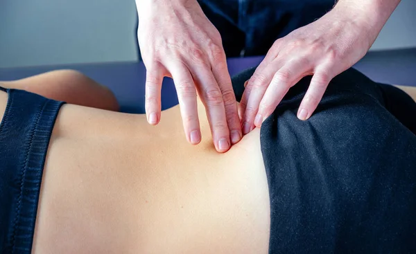
Treatment Of Sacroiliac Joint Dysfunction Or SI Joint Pain Performed By Osteopathic Doctor
Image, 3.69MB, 5863 × 3816 jpg
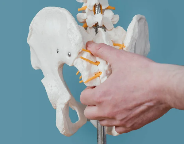
Close Up Of Male Doctors Hand Showing Sacroiliac Joint On Skeleton Spine Model
Image, 5.6MB, 4888 × 3836 jpg
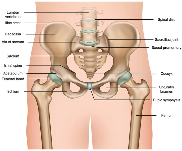
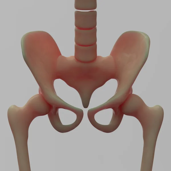
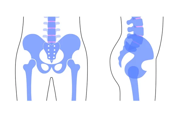
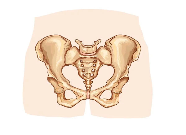
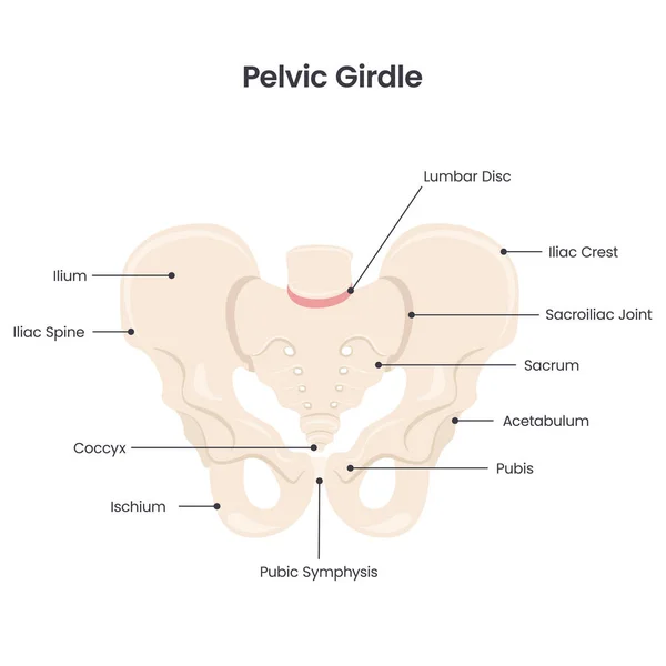
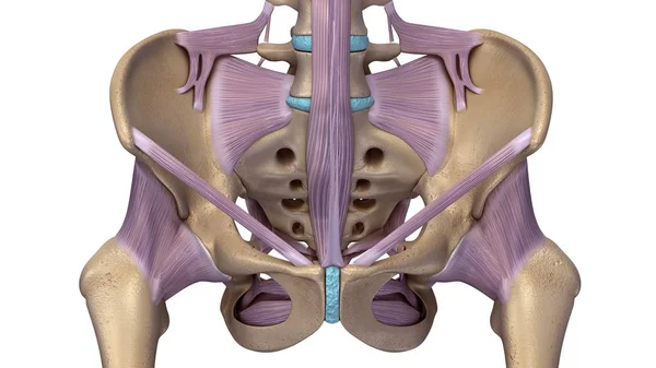
Human Pelvis Anatomy 3d Medical Vector Illustration On White Background
Vector, 5.31MB, 6000 × 5000 eps
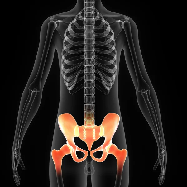


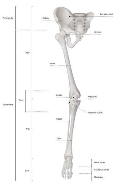
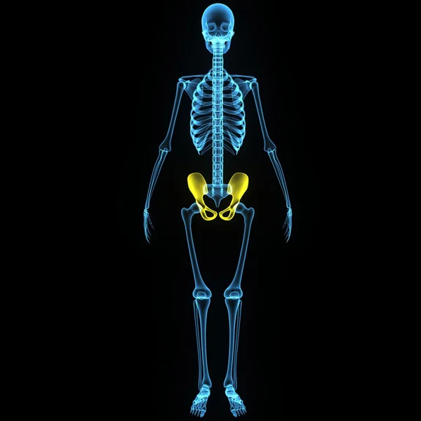
Infographic Diagram Of Human Skeleton Lower Limb Anatomy Bone System Or Leg Bone Anterior View- 3D- Human Anatomy- Medical Diagram- Educational And Human Body Concept- Isolated On White Background
Image, 6.28MB, 9405 × 15000 jpg
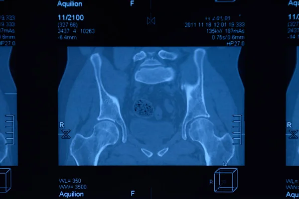
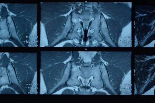
MRI Sacroiliac Articulation. Study Of Ankylosing Spondyloarthritis Patient.
Image, 8.84MB, 6000 × 4000 jpg

MRI Sacroiliac Articulation. Study Of Ankylosing Spondyloarthritis Patient.
Image, 7.17MB, 5345 × 3248 jpg
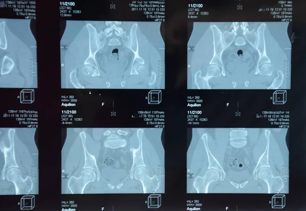
MRI Sacroiliac Articulation. Study Of Ankylosing Spondyloarthritis Patient.
Image, 9.07MB, 5372 × 3712 jpg

MRI Sacroiliac Articulation. Study Of Ankylosing Spondyloarthritis Patient.
Image, 9.96MB, 5896 × 3931 jpg


MRI Sacroiliac Articulation. Study Of Ankylosing Spondyloarthritis Patient.
Image, 9.21MB, 5782 × 3855 jpg
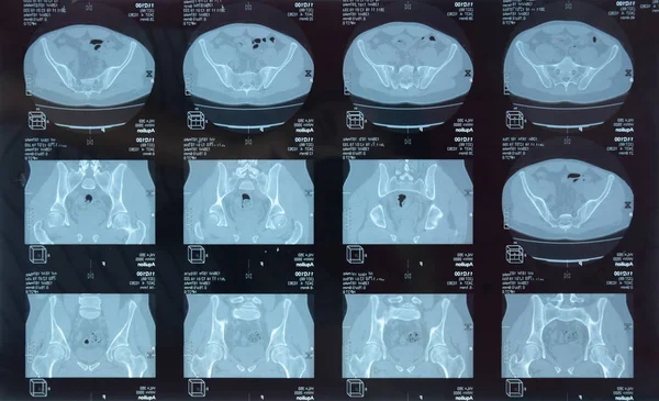
MRI Sacroiliac Articulation. Study Of Ankylosing Spondyloarthritis Patient.
Image, 8.44MB, 5791 × 3531 jpg

MRI Sacroiliac Articulation. Study Of Ankylosing Spondyloarthritis Patient.
Image, 7.86MB, 5228 × 3948 jpg
MRI Sacroiliac Articulation. Study Of Ankylosing Spondyloarthritis Patient.
Image, 8.74MB, 5942 × 3961 jpg
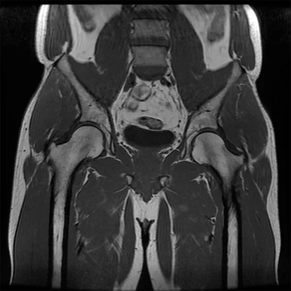
Magnetic Resonance Imaging Of The Male Hip. Observed Changes In Osteoarthritis In The Right Hip.
Image, 1.12MB, 2000 × 2000 jpg
Lumbar Spine And Sacrococcygeal On A White Background, Isolate. Intervertebral Hernia Of The Spine, Rupture Of The Fibrous Ring
Image, 0.9MB, 2959 × 3648 jpg

Magnetic Resonance Imaging Of The Male Hip - Cross Section. Observed Changes In Osteoarthritis In The Right Hip.
Image, 0.94MB, 2000 × 2000 jpg
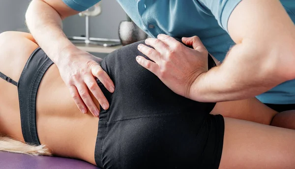
Treatment Of Sacroiliac Joint Dysfunction Or SI Joint Pain Performed By Osteopathic Doctor
Image, 2.25MB, 5185 × 2966 jpg
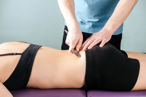
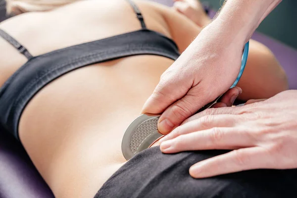
Physical Therapist Fixing Sacroiliac Joint Pain With IASTM Guasha Tool
Image, 2.4MB, 6016 × 4016 jpg


Physical Therapist Demonstrates How To Apply IASTM Tool To Treat Sacroiliac Joint Pain On Flexible Chiropractic Spine Model
Image, 3.74MB, 6016 × 4016 jpg

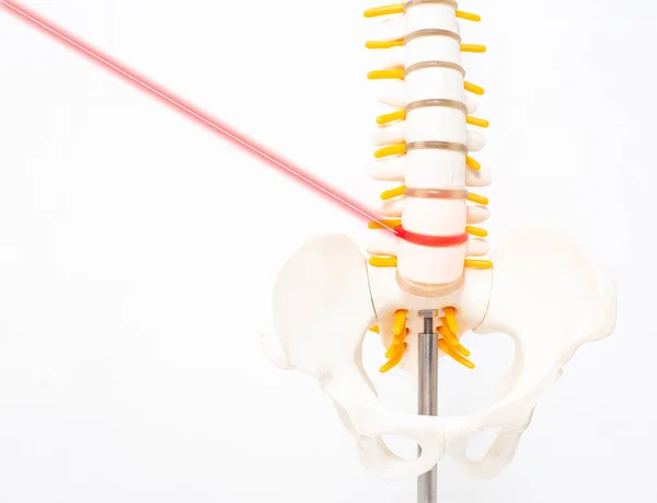
Mock Up Human Spine On A White Background With A Laser. The Concept Of A New Modern Method Without Surgery For The Treatment And Removal Of Intervertebral Hernias, Copy Space
Image, 1.15MB, 4493 × 3441 jpg
Physical Therapist Fixing Sacroiliac Joint Pain With IASTM Guasha Tool
Image, 2.77MB, 6016 × 4016 jpg
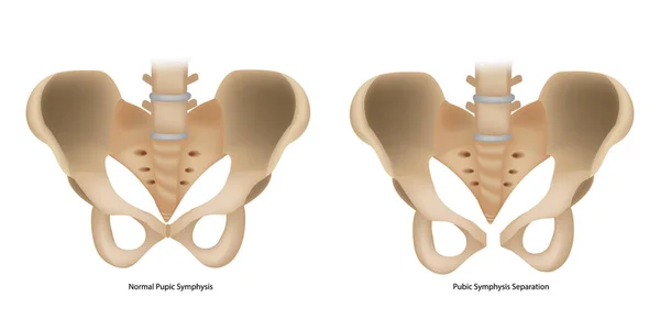
Pubic Symphysis Separation Or SPD. Symphysis Pubis Dysfunction And Normal Pupic Symphysis.
Vector, 2.2MB, 6000 × 3000 eps
Page 1 >> Next