
Stock image Sagital page 2

MRI Of The Prostate Gland Reveals Focal Abnormal SI Lesion At Left PZpl At Apex As Described; PI-RADS Category 4, Clinicall
Image, 5.38MB, 5184 × 3240 jpg


Sagittal Section Of The Right Foot In The Old Book Atlas Der Anatomie By Fischer, 1894, Jena
Image, 5.35MB, 5744 × 3240 jpg

Sagittal Section Of The Right Foot In The Old Book Atlas Der Anatomie By Fischer, 1894, Jena
Image, 5.35MB, 5744 × 3240 jpg

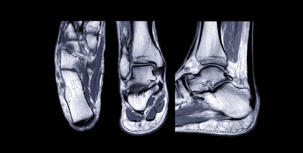
Compare MRI Ankle Joint Axial , Coronal And Sagittal T2 View For Diagnostic Tendon Injury.
Image, 3.14MB, 5076 × 2568 jpg


MRI Sacroiliac Articulation. Study Of Ankylosing Spondyloarthritis Patient.
Image, 9.21MB, 5782 × 3855 jpg
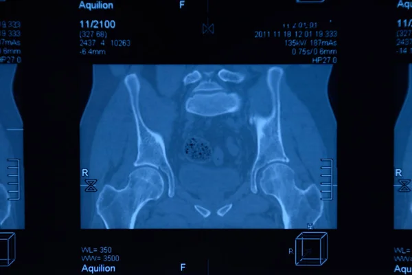
MRI Sacroiliac Articulation. Study Of Ankylosing Spondyloarthritis Patient.
Image, 8.84MB, 6000 × 4000 jpg
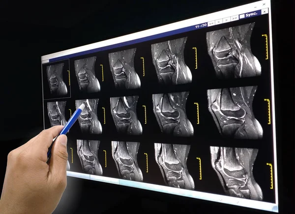
Close Up Hand Doctor Holding A Pen And Explain The Results Patient To Know Magnetic Resonance (MRI) Knee Joint.
Image, 4.3MB, 4128 × 3004 jpg

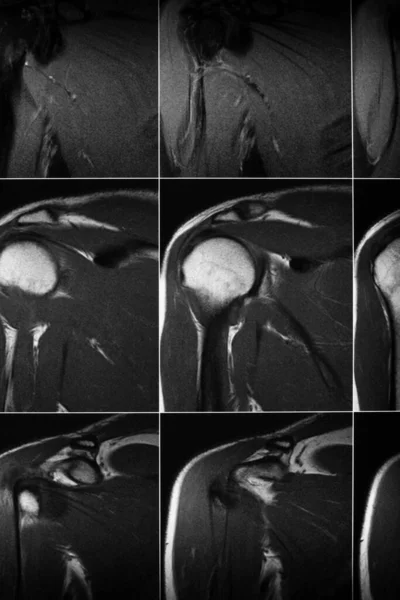
Magnetic Resonance Imaging Of Left Shoulder Rotator Cuff Tear With Suspected Lipoma Of Left Shoulder Science And Education Mri Shoulder Background,Medical.
Image, 7.69MB, 5724 × 4000 jpg
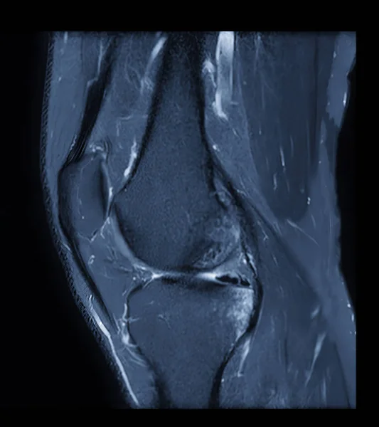
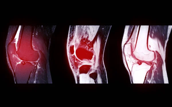
MRI Knee Or Magnetic Resonance Imaging Of Knee Joint Stir Technique Of Sagittal View For Fat Suppression.
Image, 1.96MB, 2370 × 2664 jpg
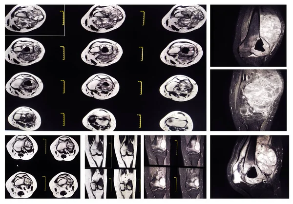
Collection MRI Knee: At Distal Lateral Femoral Metaphysis, Representing The Pre-existing Location Of The Tumor, Surrounding With A Well-defined Bony Destruction With Multiple Cystic.
Image, 8.76MB, 5672 × 3967 jpg
MRI THE BRAIN.Moderate Perilesional Vasogenic Edema With 0.7 Cm Midline Shift To The Left Side.Medical Image Concept.
Image, 2.37MB, 3000 × 3000 jpg
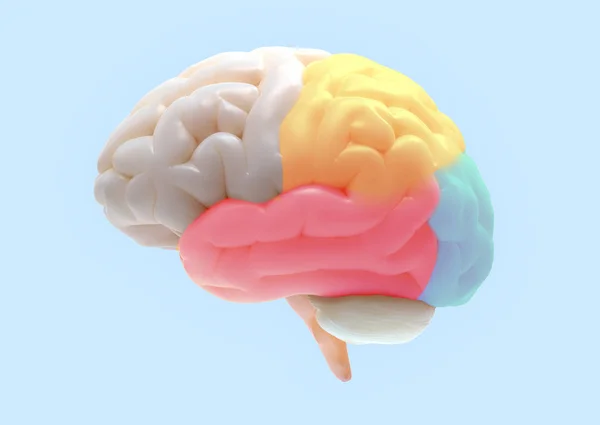
3D Brain Rendering With Subdivisions Color Parts Isolated On White Background In Soft Lighting Included Clipping Path For Use In Any Backdrop
Image, 4.73MB, 5400 × 3827 jpg

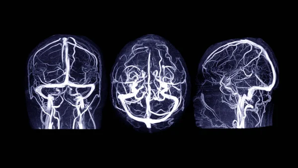
MRI OF THE BRAIN AND MRA & MRV OF THE BRAIN.Moderate Perilesional Vasogenic Edema With 0.7 Cm Midline Shift To The Left Side.
Image, 5.25MB, 8316 × 2928 jpg
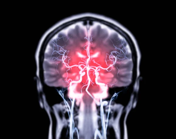
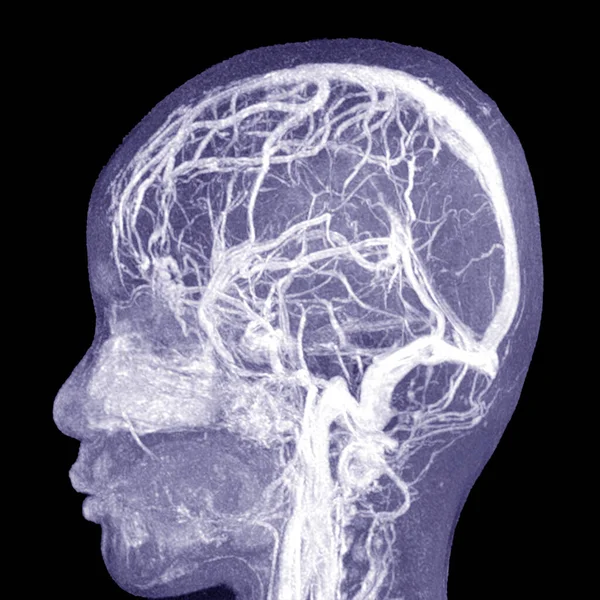
MRV Brain Or Magnetic Resonance Venography Of The Brain For Abnormalities In Venous Drainage Of The Brain
Image, 1.22MB, 3840 × 2400 jpg

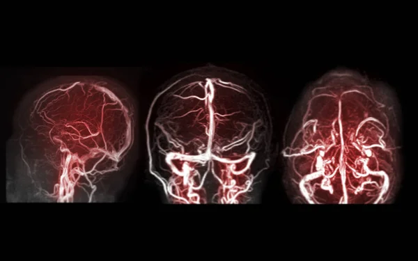
MRV Brain Or Magnetic Resonance Venography Of The Brain For Abnormalities In Venous Drainage Of The Brain
Image, 5.31MB, 6000 × 3400 jpg
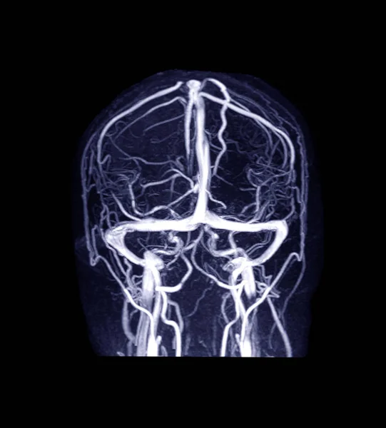
MRV Brain Or Magnetic Resonance Venography Of The Brain For Abnormalities In Venous Drainage Of The Brain
Image, 1.96MB, 2700 × 2992 jpg
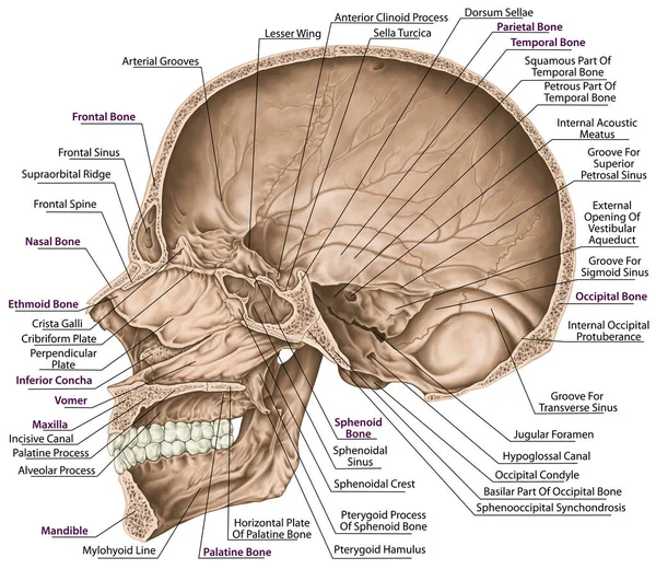
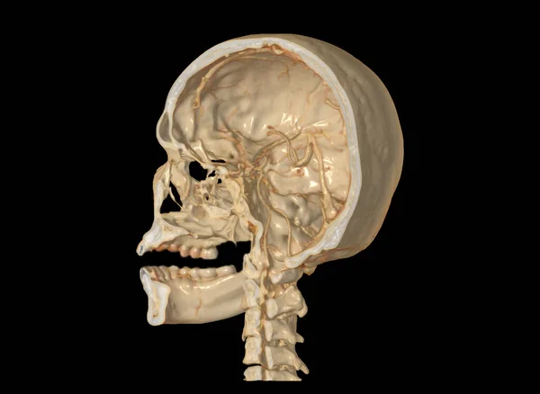
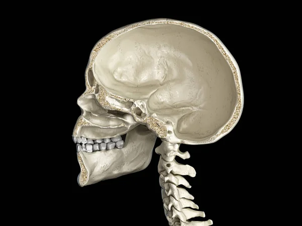
Cranial Cavity. The Bones Of The Cranium, The Bones Of The Head, Skull. Openings For Nerves And Blood Vessels, Foramens And Processes. The Names Of The Cranial Bones. Parasagittal Section.
Image, 8.84MB, 5906 × 5126 jpg
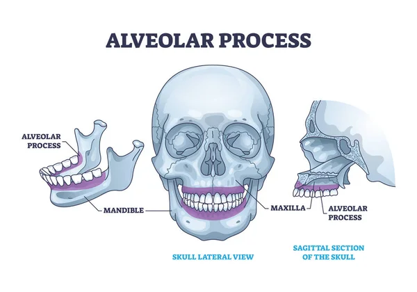
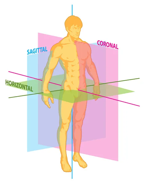
Alveolar Process With Anatomical Head Bone Ridge For Teeth Outline Diagram. Labeled Educational Scheme With Chin Maxilla And Mandible Parts Vector Illustration. Dental Implant Location On Human Skull.
Vector, 9.61MB, 5000 × 3500 eps
Compare Of MRI Knee Joint Or Magnetic Resonance Imaging Sagital View For Detect Tear Or Sprain Of The Anterior Cruciate Ligament (ACL).
Image, 2.76MB, 4448 × 2768 jpg
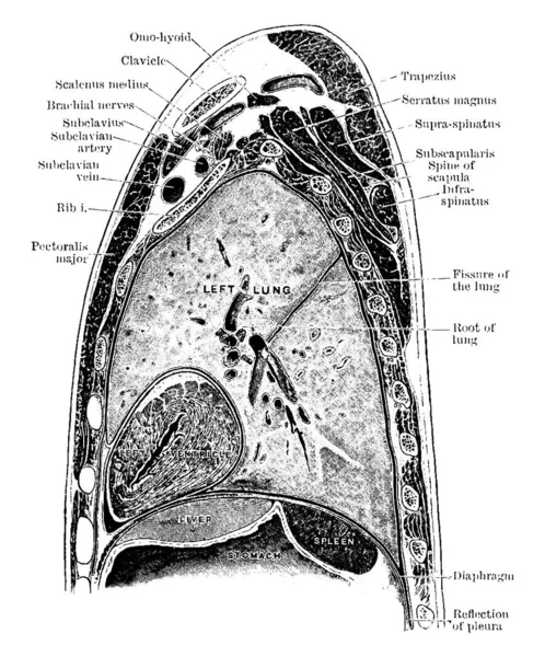
The Sagittal Section Through The Left Shoulder, Lung, And Apex Of The Heart, Vintage Line Drawing Or Engraving Illustration.
Vector, 13.91MB, 8082 × 9721 eps

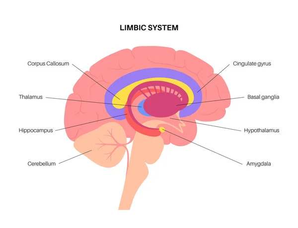
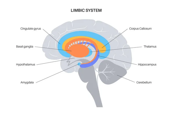
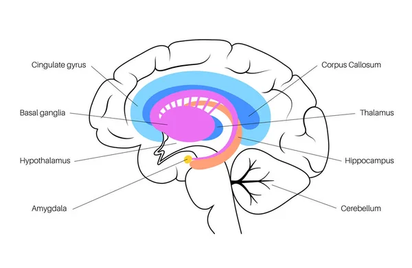
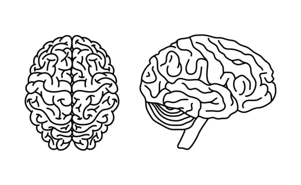
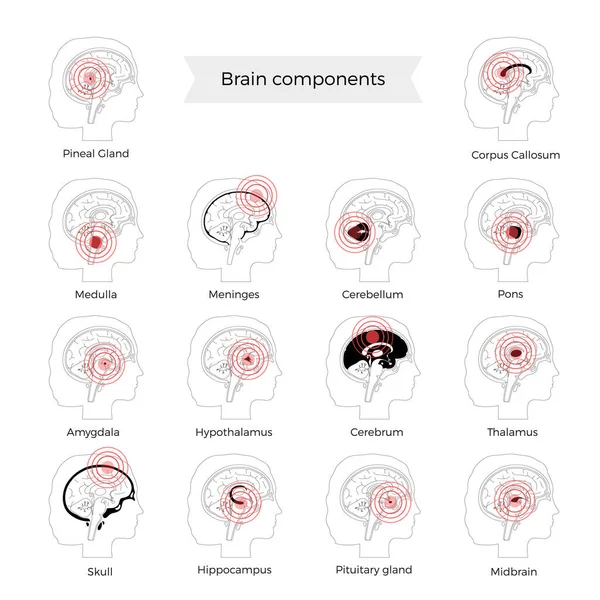
Parts Of The Brain Educational Scheme, Vector Vertical Poster Illustration On White Background
Vector, 1.3MB, 3535 × 5000 eps

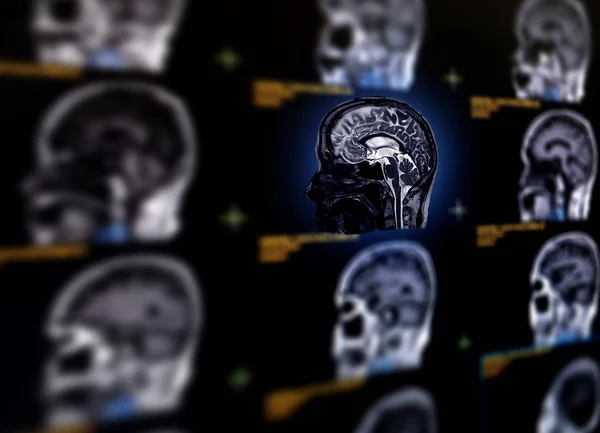
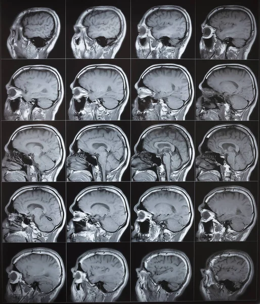
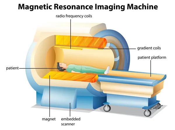
Selective Focus Of MRI Brain Sagittal Plane For Detect A Variety Of Conditions Of The Brain Such As Cysts, Tumors, Bleeding, Swelling, Developmental And Structural Abnormalities Or Infections .
Image, 7.51MB, 7584 × 5484 jpg
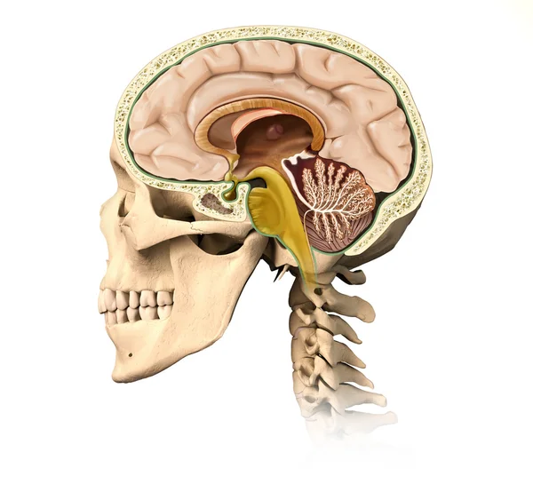
Cranial Cavity. The Bones Of The Cranium, The Bones Of The Head, Skull. Openings For Nerves And Blood Vessels, Foramens And Processes. Sagittal Section.
Image, 8.86MB, 5906 × 5906 jpg

Fontanelle And Sutures In An Infants Skull, Including Anterior And Posterior Fontanelles, Sagittal, Coronal, Lambdoidal Sutures Diagram Hand Drawn Vector Illustration. Medical Science Illustration
Vector, 0.6MB, 4000 × 4000 eps

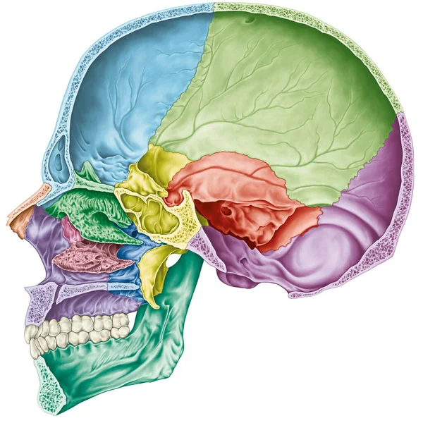
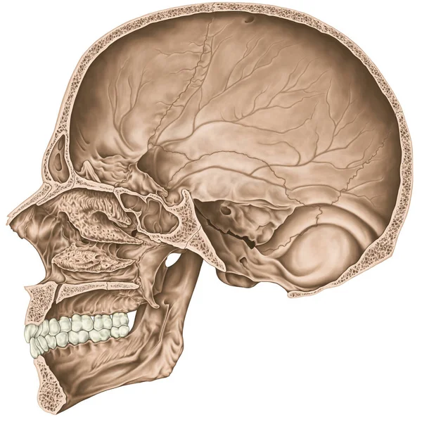
Cranial Cavity. The Bones Of The Cranium, The Bones Of The Head, Skull. The Individual Bones And Their Salient Features In Different Colors. Sagittal Section.
Image, 10.52MB, 5906 × 5906 jpg
Previous << Page 2 >> Next