
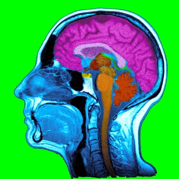
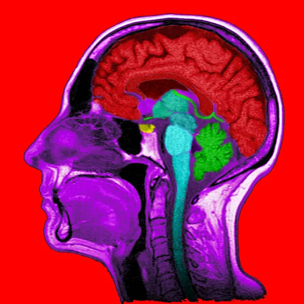
Stock image Sagittal
Collection Of CT Lumbar Or L-S Spine 3D Rendering Image Showing Compression Fractures At L2. 3D Illustration.
Image, 2.75MB, 4407 × 2680 jpg
MRI Sacroiliac Articulation. Study Of Ankylosing Spondyloarthritis Patient.
Image, 8.44MB, 5791 × 3531 jpg
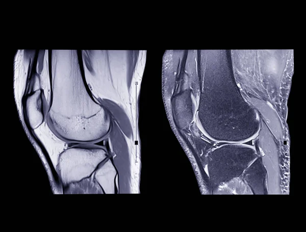
Magnetic Resonance Imaging Or MRI Knee Comparison Sagittal PDW And TIW View For Detect Tear Or Sprain Of The Anterior Cruciate Ligament (ACL).clipping Path.
Image, 2.65MB, 3968 × 3014 jpg
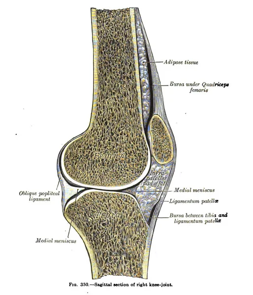
A Vertical Anatomy Drawing And Text Of The Sagittal Section Of Right Knee Joint, From The 19th Century
Image, 4.49MB, 2287 × 2666 jpg
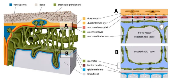
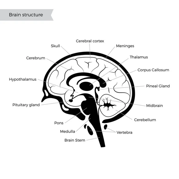
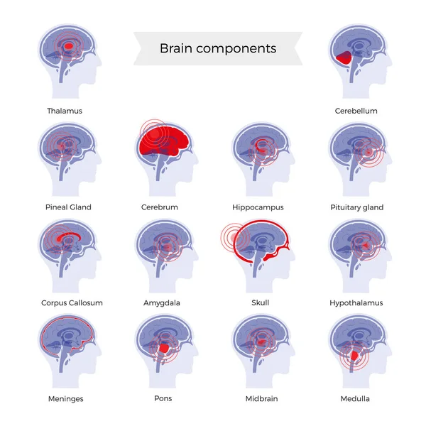
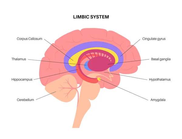
Protective Membranes Covering The Brain. Meninges: Dura Mater, Arachnoid, And Pia Mater. Cross Section Of The Human Brain. Layers. Diagram For Educational, Medical, Biological, Scientific Usem 3d Render
Image, 4MB, 5335 × 2599 jpg

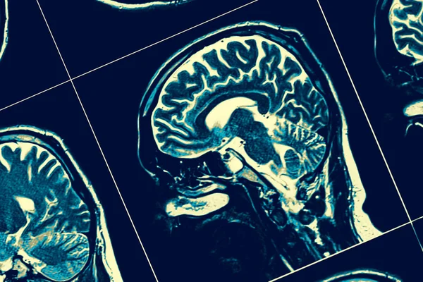
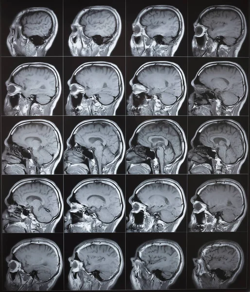
Selective Focus Of MRI Brain Sagittal Plane For Detect A Variety Of Conditions Of The Brain Such As Cysts, Tumors, Bleeding, Swelling, Developmental And Structural Abnormalities Or Infections .
Image, 6.81MB, 7056 × 5208 jpg


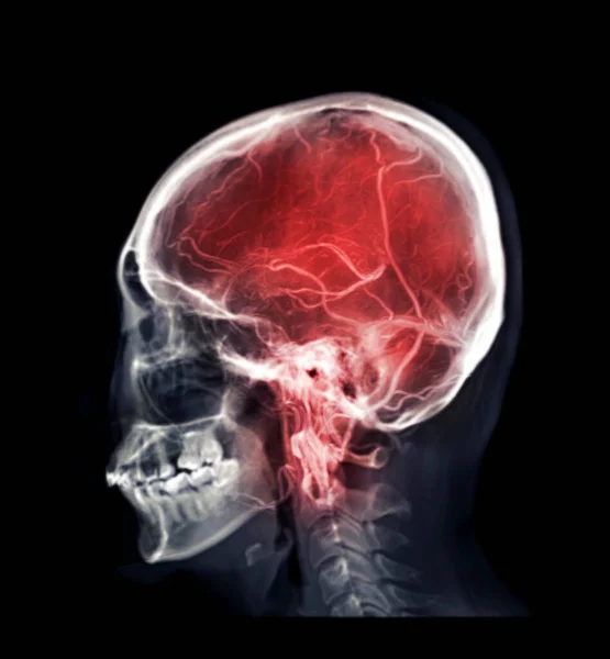
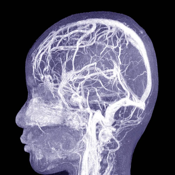
Mix Skull Image And MRV Brain Or Magnetic Resonance Venography Of The Brain For Abnormalities In Venous Drainage Of The Brain
Image, 1.32MB, 2999 × 3240 jpg

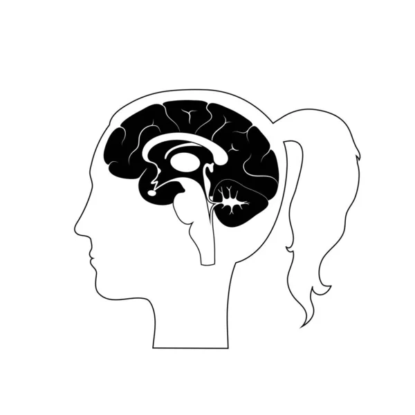
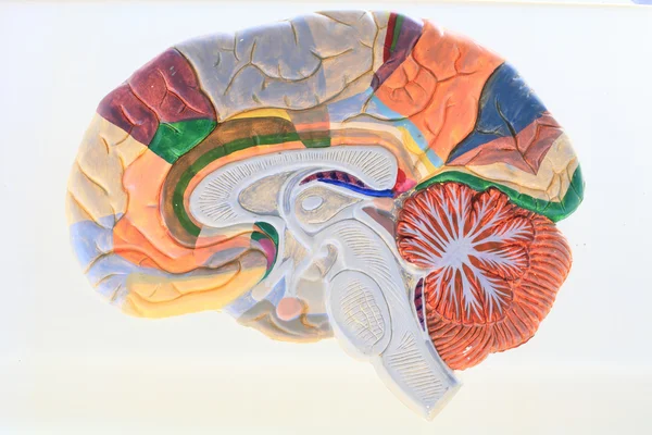
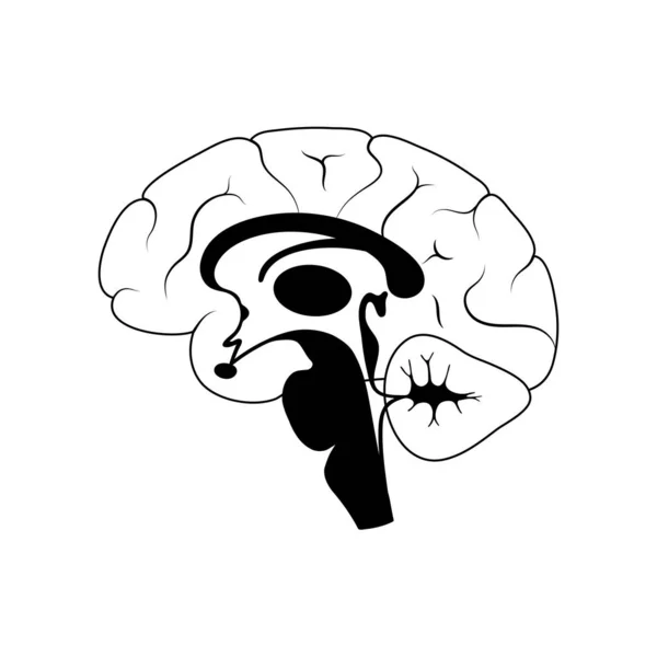
Sagittal Section Of The Brain With Meninges And Cerebrospinal Fluid. .
Image, 0.84MB, 3630 × 3114 jpg
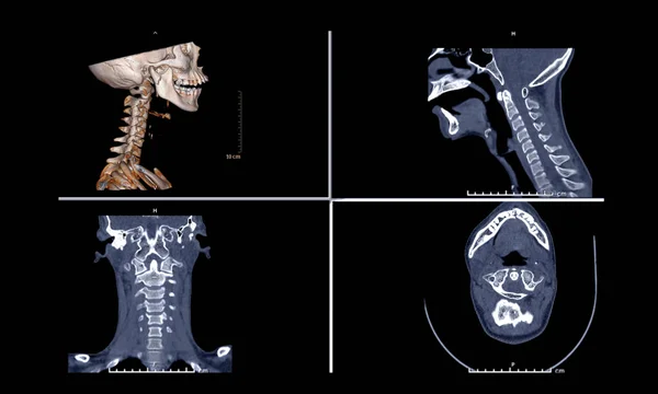
Comparison Of CT C-Spine Or Cervical Spine 3D Rendering Image , Sagittal ,Corona And Axiall View In Patient Trauma Head Injury.
Image, 2.06MB, 5008 × 3008 jpg
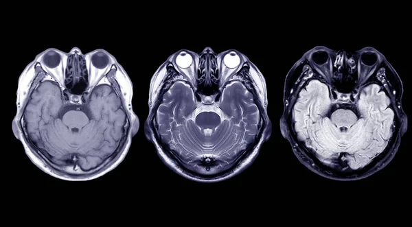
Compare MRI Of The Brain Axial T1, T2 And T2 Flair View For Detect A Variety Of Conditions Of The Brain Such As Cysts, Tumors, Bleeding Isolated On Black Backgroud , Bleeding .
Image, 2.85MB, 4912 × 2712 jpg
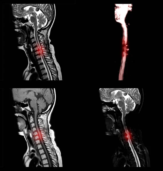
A Sagittal View Of MRI C-spine Or Magnetic Resonance Image Of Cervical Spine Showing Spondylosis Causing Cervical Spondylotic Myelopathy And Compression Fracture.
Image, 1.46MB, 2835 × 2976 jpg

Selective Focus Of MRI Brain Sagittal Plane For Detect A Variety Of Conditions Of The Brain Such As Tumors. Idea Concept.
Image, 7.63MB, 6960 × 5256 jpg


MRI Of The Prostate Gland Reveals Focal Abnormal SI Lesion At Left PZpl At Apex As Described; PI-RADS Category 4, Clinicall
Image, 5.38MB, 5184 × 3240 jpg


Sagittal Section Of The Right Foot In The Old Book Atlas Der Anatomie By Fischer, 1894, Jena
Image, 5.35MB, 5744 × 3240 jpg

Collection Transparent Image Of The Skull Blue Color With MRI Brain For Medical Background Concept.
Image, 5.96MB, 6544 × 4112 jpg


Sagittal Section Of The Right Foot In The Old Book Atlas Der Anatomie By Fischer, 1894, Jena
Image, 5.35MB, 5744 × 3240 jpg

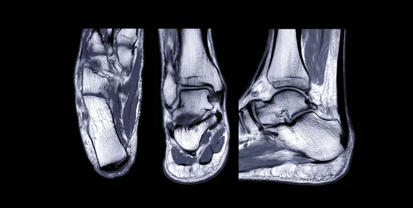
Compare MRI Ankle Joint Axial , Coronal And Sagittal T2 View For Diagnostic Tendon Injury.
Image, 3.14MB, 5076 × 2568 jpg


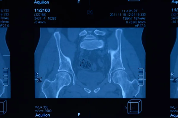
MRI Sacroiliac Articulation. Study Of Ankylosing Spondyloarthritis Patient.
Image, 9.21MB, 5782 × 3855 jpg
MRI Sacroiliac Articulation. Study Of Ankylosing Spondyloarthritis Patient.
Image, 8.84MB, 6000 × 4000 jpg
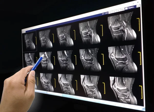
Close Up Hand Doctor Holding A Pen And Explain The Results Patient To Know Magnetic Resonance (MRI) Knee Joint.
Image, 4.3MB, 4128 × 3004 jpg

Magnetic Resonance Imaging Of Left Shoulder Rotator Cuff Tear With Suspected Lipoma Of Left Shoulder Science And Education Mri Shoulder Background,Medical.
Image, 7.69MB, 5724 × 4000 jpg
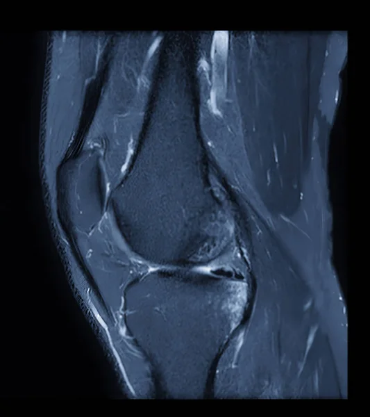
MRI Knee Or Magnetic Resonance Imaging Of Knee Joint Stir Technique Of Sagittal View For Fat Suppression.
Image, 1.96MB, 2370 × 2664 jpg
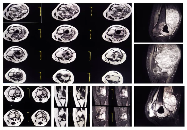
Collection MRI Knee: At Distal Lateral Femoral Metaphysis, Representing The Pre-existing Location Of The Tumor, Surrounding With A Well-defined Bony Destruction With Multiple Cystic.
Image, 8.76MB, 5672 × 3967 jpg
MRI THE BRAIN.Moderate Perilesional Vasogenic Edema With 0.7 Cm Midline Shift To The Left Side.Medical Image Concept.
Image, 2.37MB, 3000 × 3000 jpg

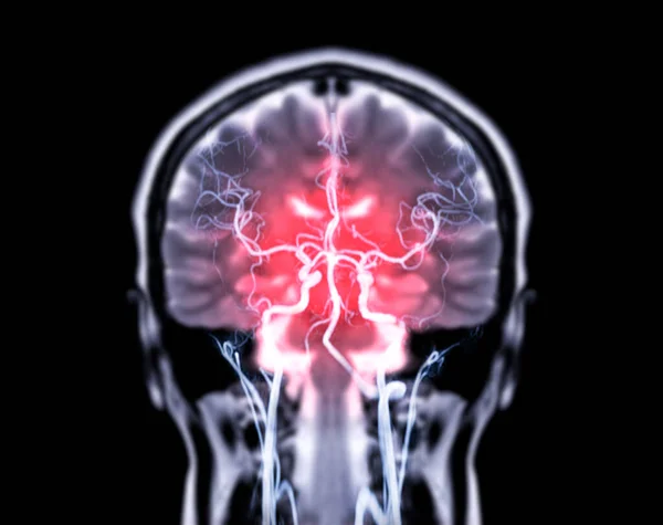
MRI OF THE BRAIN AND MRA & MRV OF THE BRAIN.Moderate Perilesional Vasogenic Edema With 0.7 Cm Midline Shift To The Left Side.
Image, 5.25MB, 8316 × 2928 jpg
Page 1 >> Next