
Stock image Sagittal page 3
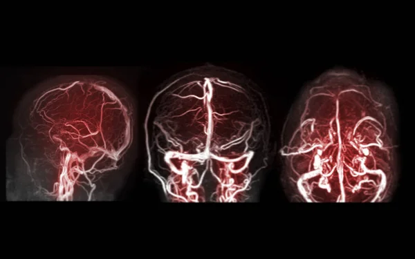
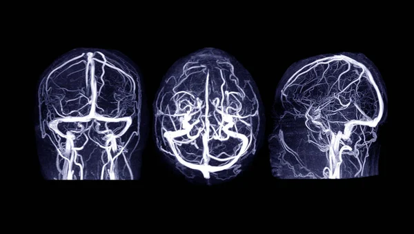
MRV Brain Or Magnetic Resonance Venography Of The Brain For Abnormalities In Venous Drainage Of The Brain
Image, 1.22MB, 3840 × 2400 jpg

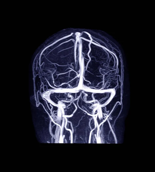
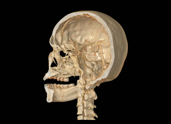
MRV Brain Or Magnetic Resonance Venography Of The Brain For Abnormalities In Venous Drainage Of The Brain
Image, 1.96MB, 2700 × 2992 jpg
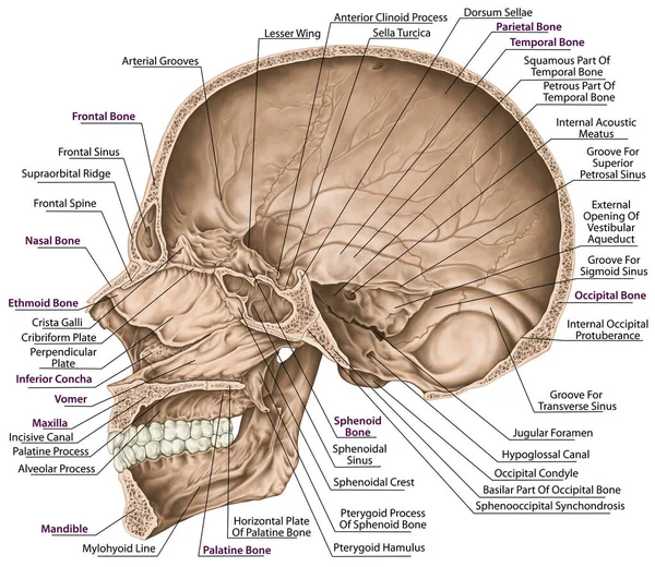
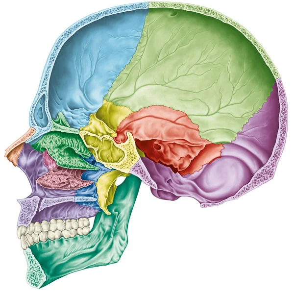
Cranial Cavity. The Bones Of The Cranium, The Bones Of The Head, Skull. Openings For Nerves And Blood Vessels, Foramens And Processes. The Names Of The Cranial Bones. Parasagittal Section.
Image, 8.84MB, 5906 × 5126 jpg
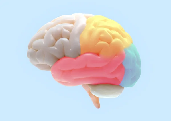
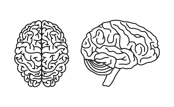
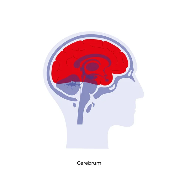
3D Brain Rendering With Subdivisions Color Parts Isolated On White Background In Soft Lighting Included Clipping Path For Use In Any Backdrop
Image, 4.73MB, 5400 × 3827 jpg
MRV Brain Or Magnetic Resonance Venography Of The Brain For Abnormalities In Venous Drainage Of The Brain
Image, 5.31MB, 6000 × 3400 jpg
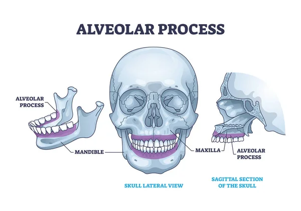
Alveolar Process With Anatomical Head Bone Ridge For Teeth Outline Diagram. Labeled Educational Scheme With Chin Maxilla And Mandible Parts Vector Illustration. Dental Implant Location On Human Skull.
Vector, 9.61MB, 5000 × 3500 eps
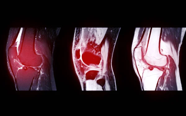
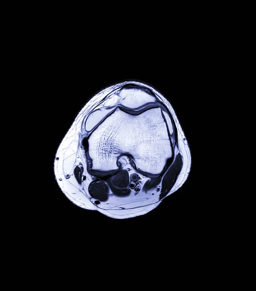
Compare Of MRI Knee Joint Or Magnetic Resonance Imaging Sagital View For Detect Tear Or Sprain Of The Anterior Cruciate Ligament (ACL).
Image, 2.76MB, 4448 × 2768 jpg
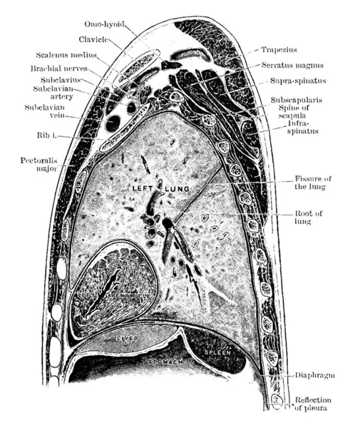
The Sagittal Section Through The Left Shoulder, Lung, And Apex Of The Heart, Vintage Line Drawing Or Engraving Illustration.
Vector, 13.91MB, 8082 × 9721 eps

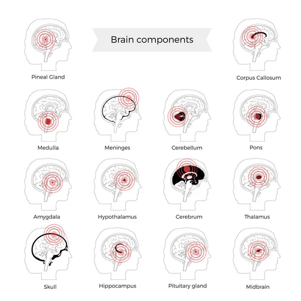
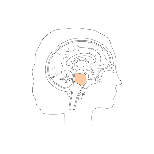
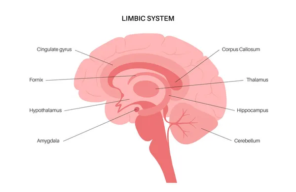
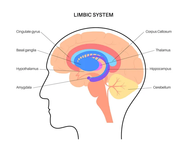
Parts Of The Brain Educational Scheme, Vector Vertical Poster Illustration On White Background
Vector, 1.3MB, 3535 × 5000 eps

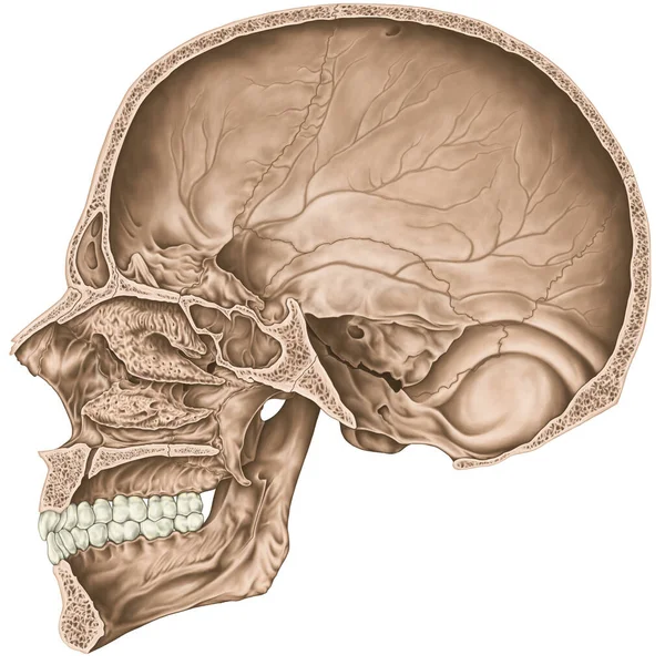
Cranial Cavity. The Bones Of The Cranium, The Bones Of The Head, Skull. Openings For Nerves And Blood Vessels, Foramens And Processes. Sagittal Section.
Image, 8.86MB, 5906 × 5906 jpg

Fontanelle And Sutures In An Infants Skull, Including Anterior And Posterior Fontanelles, Sagittal, Coronal, Lambdoidal Sutures Diagram Hand Drawn Vector Illustration. Medical Science Illustration
Vector, 0.6MB, 4000 × 4000 eps

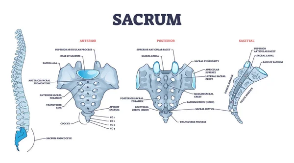
Sacrum As Spinal Bone Structure Anatomical Description Outline Diagram
Vector, 9.98MB, 6000 × 3333 eps
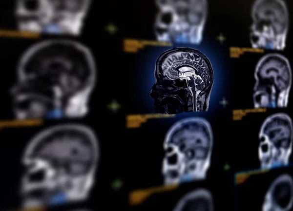
Selective Focus Of MRI Brain Sagittal Plane For Detect A Variety Of Conditions Of The Brain Such As Cysts, Tumors, Bleeding, Swelling, Developmental And Structural Abnormalities Or Infections .
Image, 7.51MB, 7584 × 5484 jpg
Cranial Cavity. The Bones Of The Cranium, The Bones Of The Head, Skull. The Individual Bones And Their Salient Features In Different Colors. Sagittal Section.
Image, 10.52MB, 5906 × 5906 jpg
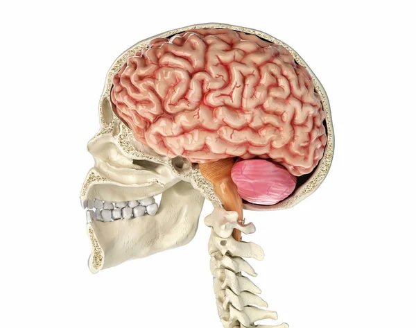
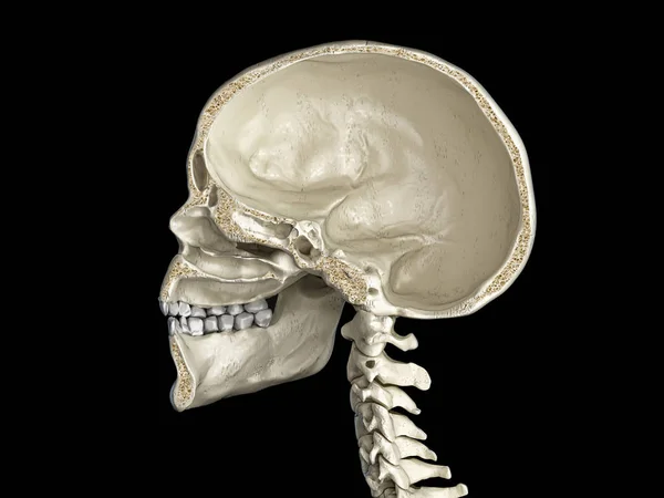
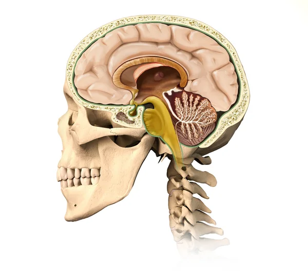
Human Skull Mid Sagittal Cross-section With Brain. Side View On Black Background.
Image, 0MB, 8234 × 6500 jpg
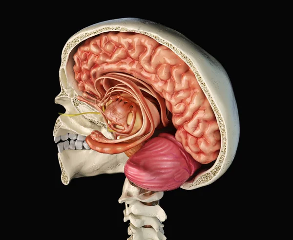
Human Skull Mid Sagittal Cross-section With Brain. Perspective View On Black Background.
Image, 0MB, 8314 × 6803 jpg


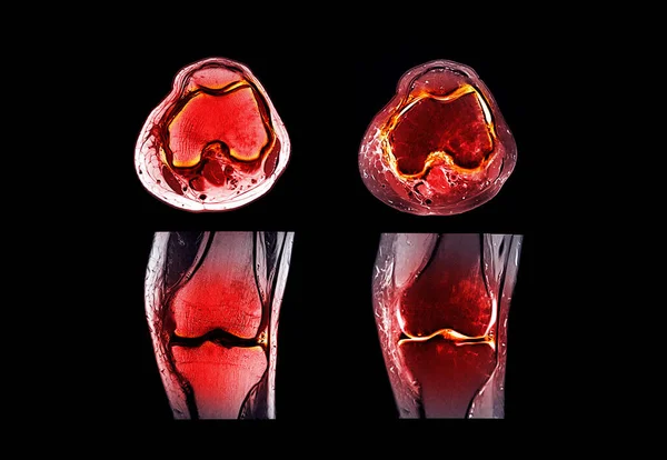
Magnetic Resonance Imaging Or MRI Knee Joint Comparison Coronal And Sagittal View For Detect Tear Or Sprain Of The Anterior Cruciate Ligament (ACL)
Image, 1.92MB, 3149 × 2173 jpg

Parts Of The Brain Landing Page Template. Tiny Male Character Balancing On Rope At Huge Human Brain Anatomy
Vector, 1.94MB, 8331 × 5000 eps
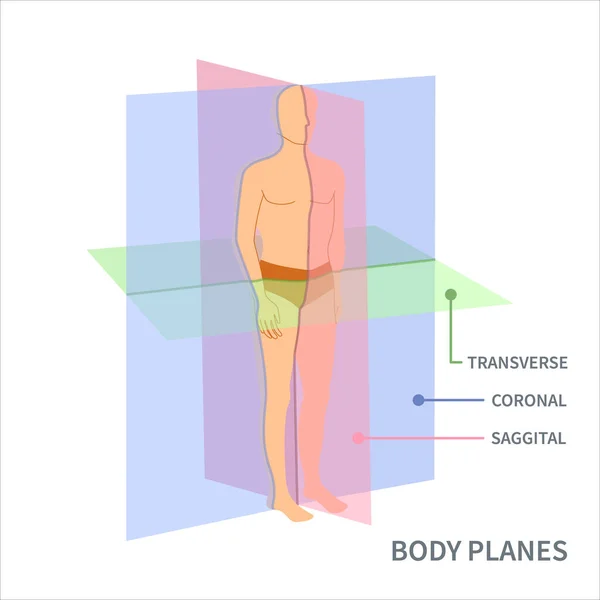
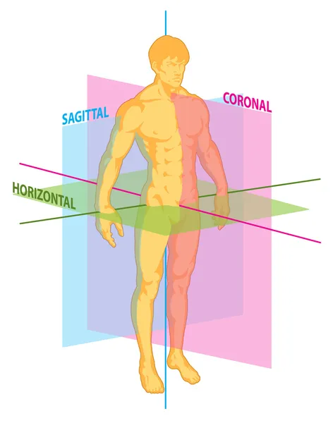
Body Anatomical Position Diagram. Sagittal, Coronal And Transverse Scanning Plane Types Shown On A Male Body. Medical Concept. Vector Illustration.
Vector, 0.35MB, 4500 × 4500 eps
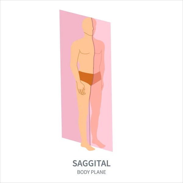
Sagittal Scanning Plane Shown On A Male Body. Human Body Anatomical Position Diagram. Probe Orientation Infographics. Medical Sonography Concept. Vector Illustration.
Vector, 0.28MB, 4500 × 4500 eps
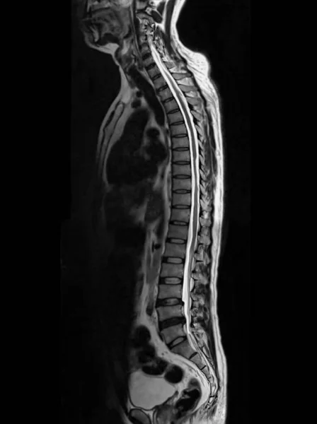
MRI Of Whole Spine T2W Sagittal Plane For Diagnostic Spinal Cord Compression.
Image, 1.94MB, 3088 × 4136 jpg

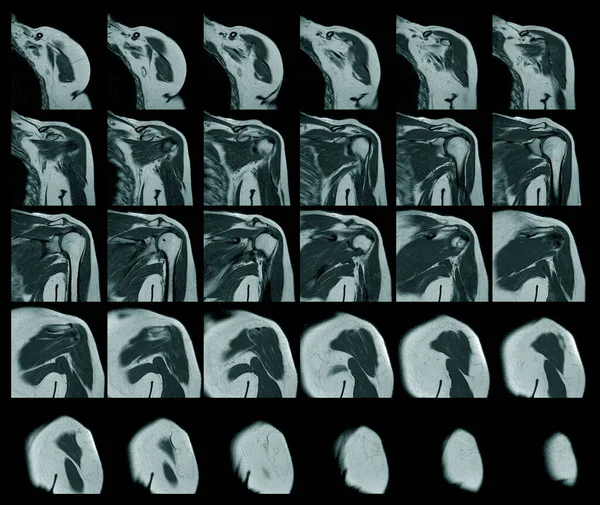
Magnetic Resonance Imaging Of Left Shoulder Rotator Cuff Tear With Suspected Lipoma Of Left Shoulder Science And Education Mri Shoulder Background,Medical.
Image, 10.26MB, 6408 × 5403 jpg

Parts Of The Brain Educational Scheme, Vector Horizontal Banner Illustration On White Background
Vector, 1.04MB, 5000 × 3330 eps
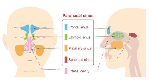
Illustrative Illustrations Of The Anatomy Of The Paranasal Sinuses From Frontal And Lateral Sagittal Plane Views
Vector, 5.41MB, 5500 × 2962 eps

MRI Of The Whole Spine Reveals Detailed Images Of The Spinal Cord For Comprehensive Evaluation.
Image, 4.48MB, 3536 × 7369 jpg

CT Knee Joint 3D Rendering Image Lateral View And Sagittal View Isolated On Black Background Showing Fracture Femur Bone.
Image, 2.15MB, 3840 × 2400 jpg

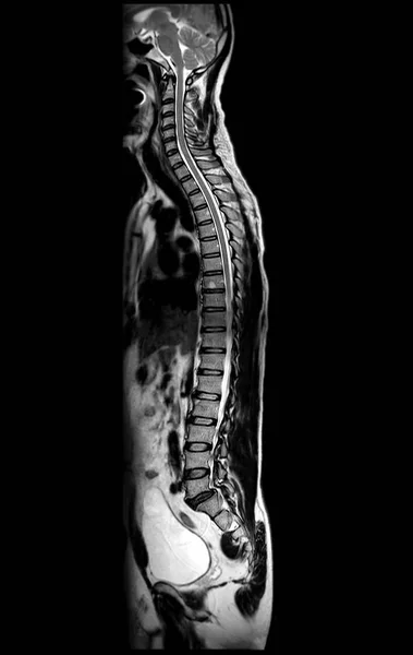
MRI Of Whole Spine T2W Sagittal Plane For Diagnostic Spinal Cord Compression.
Image, 4.7MB, 4624 × 7314 jpg
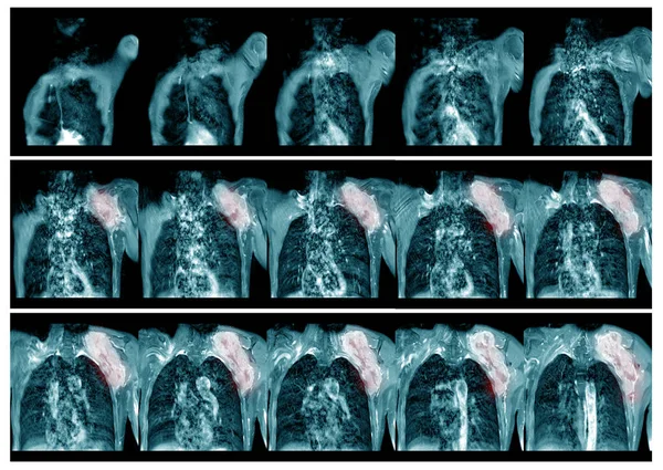
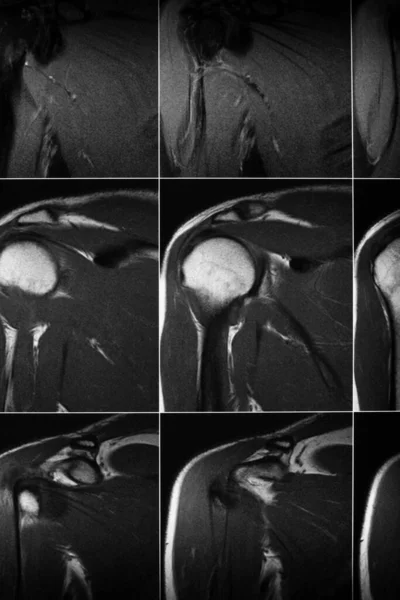
Magnetic Resonance Imaging(MRI) Left.shoulder History:Case Lt. Shoulder Mass . Impression:-The Mass Soft Tissue Sarcoma And Metastasis.Medical And Healthcare Concept.
Image, 16.6MB, 8552 × 6068 jpg
Previous << Page 3 >> Next