
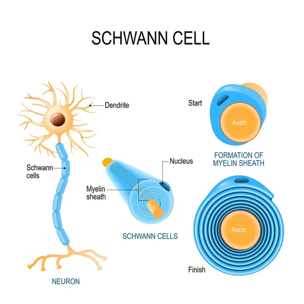
Stock image Schwann page 2
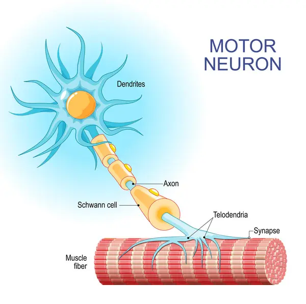
Motor Neuron. Structure And Anatomy Of A Efferent Neuron. Close-up Of A Muscle Fiber, And Motoneuron With Dendrites, Synapse, Telodendria, Axon, Schwann Cell. The Axons Carry Signals From The Spinal Cord To Muscles. Vector Illustration
Vector, 5.05MB, 4444 × 4422 eps
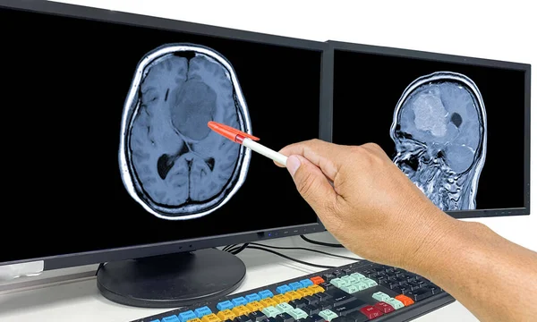
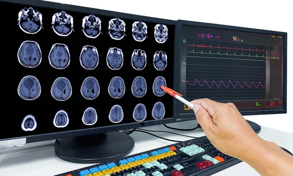
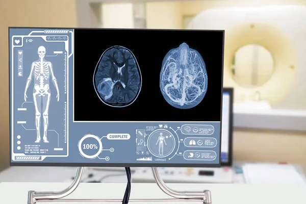
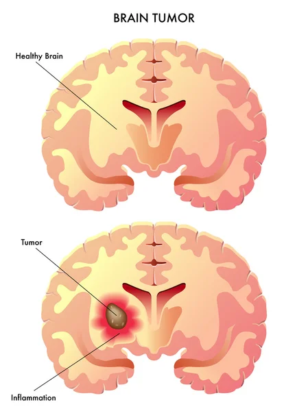
Glioblastoma, Brain Metastasis,MRI Brain The Doctor Pointed Out The Location Of The Brain Tumor On The Computer Screen
Image, 9.74MB, 7000 × 4205 jpg
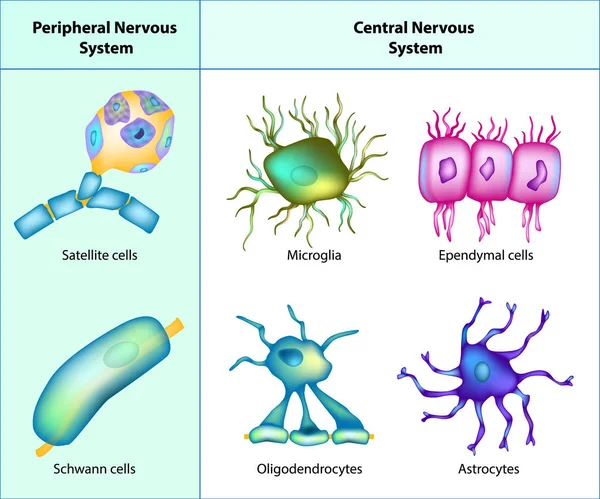
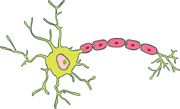
Types Of Neuroglia: Oligodendrocytes, Astrocytes, Microglia, Schwann Cells, Satellite Cells, Ependymal Cells.
Vector, 4.54MB, 5000 × 4165 eps

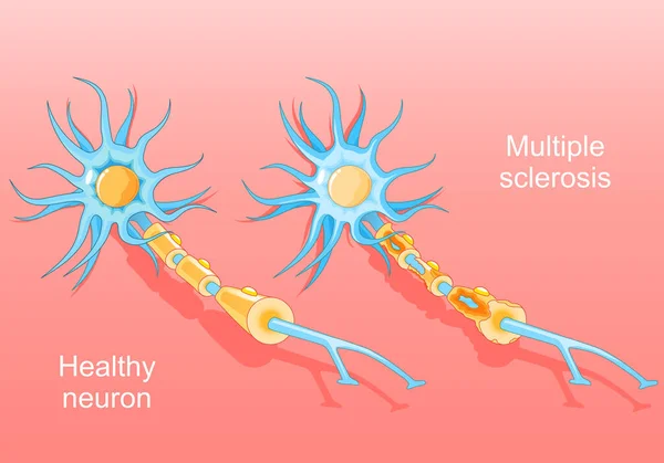
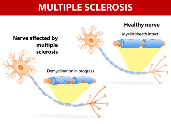
Multiple Sclerosis. Autoimmune Disease. Normal Neuron And A Neuron With A Damaged Myelin Sheath. Nerve Damage. Isometric Vector. Flat Illustration
Vector, 1.07MB, 5000 × 3498 eps
Glioblastoma, Brain Metastasis,MRI Brain The Doctor Pointed Out The Location Of The Brain Tumor On The Computer Screen
Image, 4.5MB, 7000 × 4205 jpg

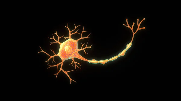
3d Rendering Of Multipolar Neurons. Multipolar Neurons Are The Most Common Type Of Neuron
Image, 0.57MB, 3840 × 2160 jpg

3d Rendering Of Multipolar Neurons. Multipolar Neurons Are The Most Common Type Of Neuron
Image, 0.47MB, 3840 × 2160 jpg

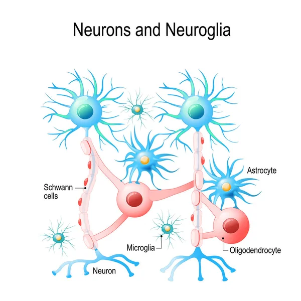
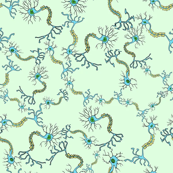
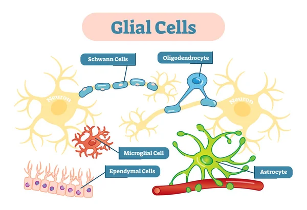
Neurons And Neuroglial Cells. Glial Cells Are Non-neuronal Cells In Brain. There Are Different Types Of Glial Cells: Oligodendrocyte, Microglia, Astrocytes And Schwann Cells. Vector Diagram For Educational, Medical, Biological And Science Use
Vector, 5.27MB, 4808 × 4808 eps

3d Rendering Of Multipolar Neurons. Multipolar Neurons Are The Most Common Type Of Neuron
Image, 0.27MB, 3840 × 2160 jpg

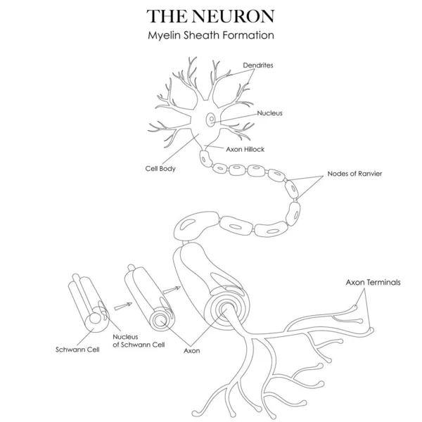
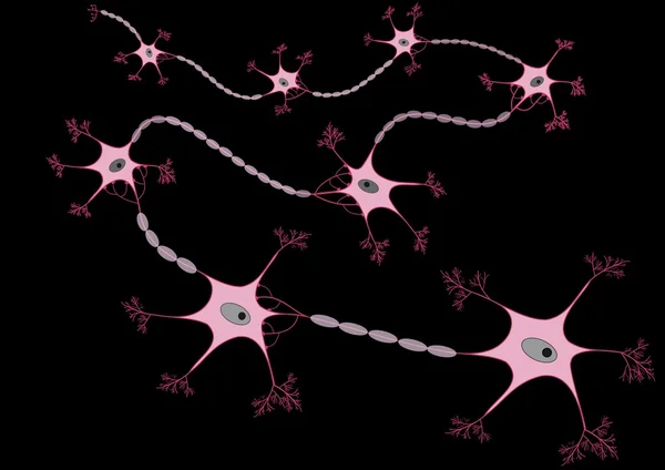
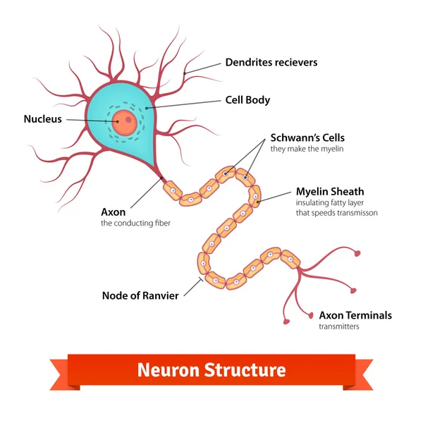
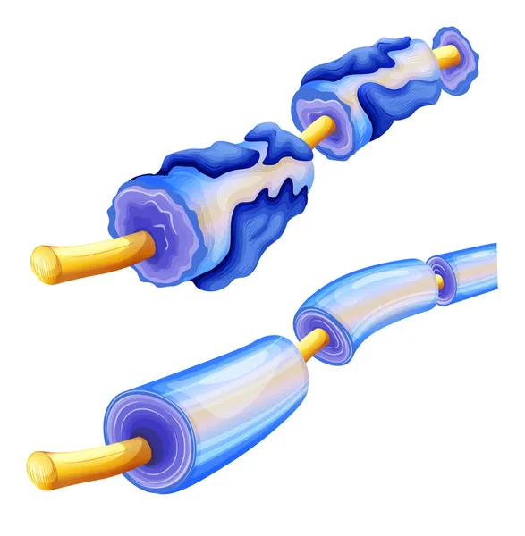
Neuron Anatomy And Myelin Sheath Formation Black And White Line Art Illustration. Can Be Used As A Worksheet For Coloring And Learning Neuron Structure
Vector, 0.65MB, 5000 × 5000 eps

3d Rendering Of Multipolar Neurons. Multipolar Neurons Are The Most Common Type Of Neuron
Image, 0.8MB, 3840 × 2160 jpg

3d Rendering Of Multipolar Neurons. Multipolar Neurons Are The Most Common Type Of Neuron
Image, 0.49MB, 3840 × 2160 jpg
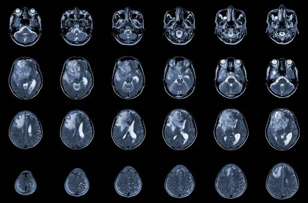
MRI Brain Axial Views .to Evaluate Brain Tumor. Glioblastoma, Brain Metastasis Isodensity Mass With An Ill-defined Margin And Surrounding Edema At The Right Frontal Lobe.
Image, 5.97MB, 6000 × 3976 jpg

Multiple Sclerosis Anatomical Vector Illustration Diagram, Medical Scheme.
Vector, 5.14MB, 5676 × 5399 eps
MRI OF THE BRAIN AND MRA & MRV OF THE BRAIN Acute Parenchymal Hemorrhage With Small Portion Of Subacute Stage Centered Medical Healthcare And Technology Concept.
Image, 4.67MB, 6000 × 4000 jpg

3d Rendering Of Multipolar Neurons. Multipolar Neurons Are The Most Common Type Of Neuron
Image, 0.12MB, 3840 × 2160 jpg
Glioblastoma Multiforme (GBM) Brain Cancer - Isometric View 3d Illustration
Image, 7.4MB, 10000 × 6600 jpg

An Interesting Photo Taken With A Microscope. Unmyelinated Fibers In Peripheral Nerves. Longitudinal Section. Hematoxylin And Eosin Stainit.
Image, 2.42MB, 3000 × 2248 jpg
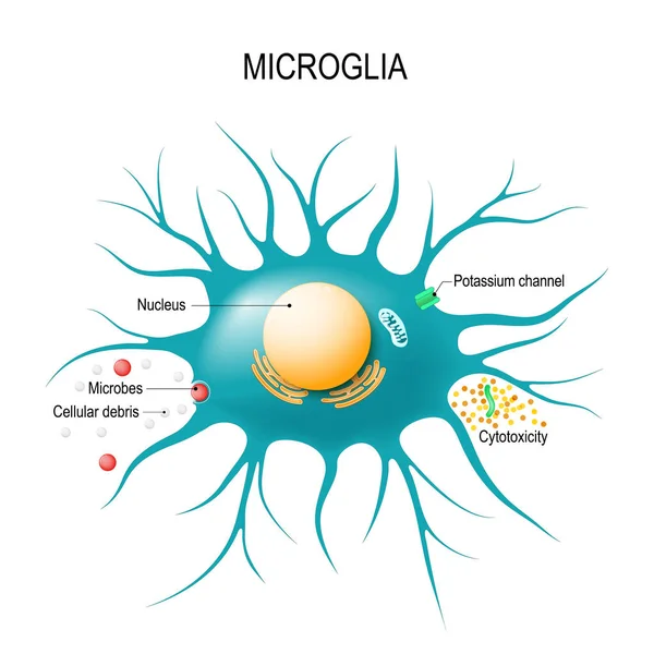
Anatomy Of A Microglial Cell. Glial Cell Is The Macrophage For Immune Defence The Central Nervous System. Vector Diagram For Educational, Medical, Biological And Science Use
Vector, 2.19MB, 5478 × 5479 eps

Close-up Of A Patient's Back Showing Multiple Neurofibromatosis Tumors On The Skin
Image, 14.92MB, 5376 × 3584 jpg

Close-up Of A Patient's Back Showing Multiple Neurofibromatosis Tumors On The Skin
Image, 14.92MB, 5376 × 3584 jpg

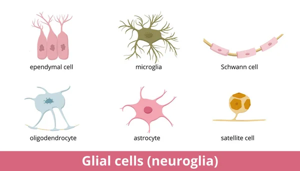
Glial Cells (neuroglia). Six Types Of Gliocytes In The Central And The Peripheral Nervous System: Oligodendrocyte, Astrocyte, Ependymal Cell, Microglia, Schwann And Satellite Cell.
Vector, 6.07MB, 7292 × 4167 eps
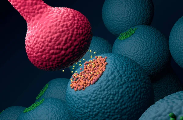
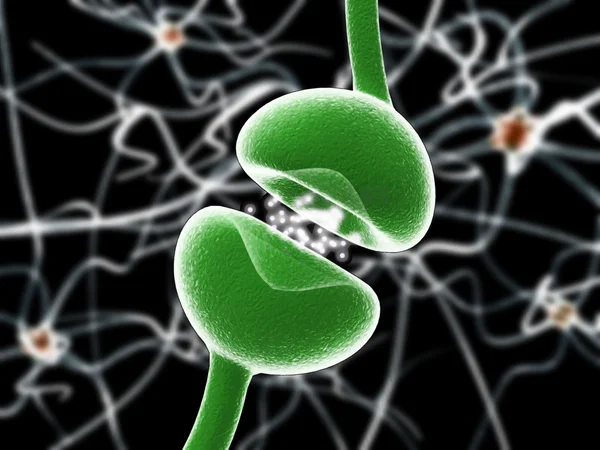
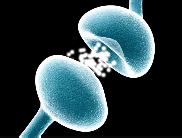
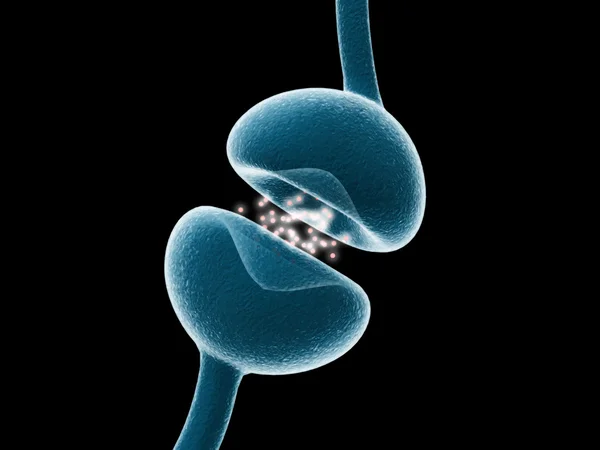
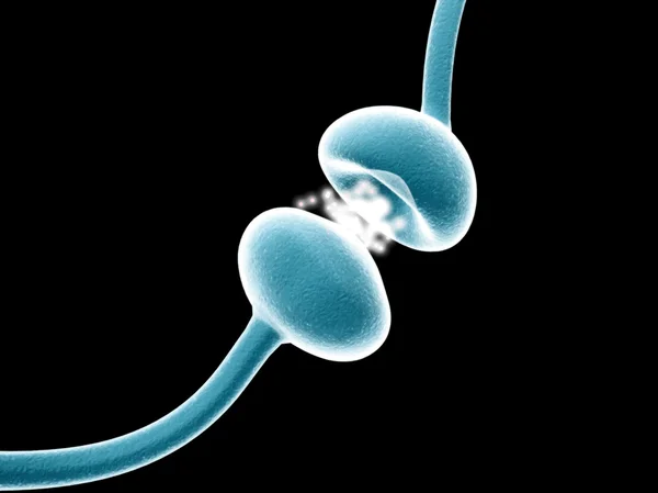
Postsynaptic Neuron Receptors (dendrite) Receive Emitted Neurotransmitters From Presynaptic Neuron (axon) - Isometric View 3d Illustration
Image, 8.03MB, 10000 × 6600 jpg

3d Rendering Of Multipolar Neurons. Multipolar Neurons Are The Most Common Type Of Neuron
Image, 0.21MB, 3840 × 2160 jpg

3d Rendering Of Multipolar Neurons. Multipolar Neurons Are The Most Common Type Of Neuron
Image, 0.73MB, 3840 × 2160 jpg

3d Rendering Of Multipolar Neurons. Multipolar Neurons Are The Most Common Type Of Neuron
Image, 0.75MB, 3840 × 2160 jpg


Previous << Page 2 >> Next