
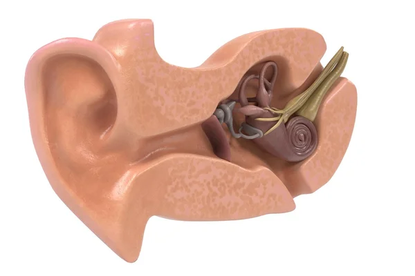
Stock image Semicircular Canals
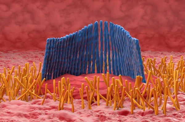
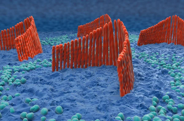
Inner Ear Hair Cell In The Vestibular System - Closeup View 3d Illustration
Image, 6.89MB, 10000 × 6600 jpg
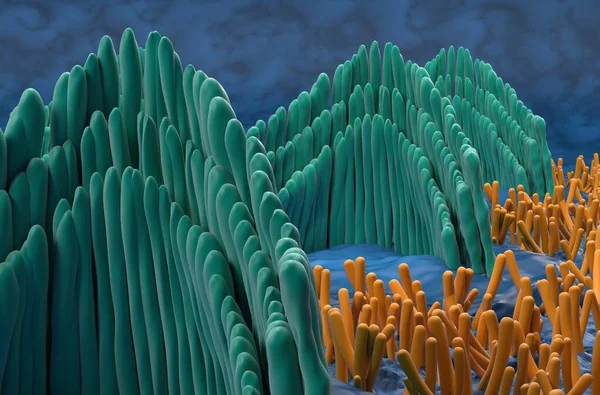
Inner Ear Hair Cells In The Vestibular System - Closeup View 3d Illustration
Image, 6.39MB, 10000 × 6600 jpg
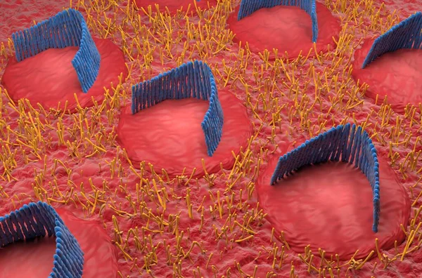
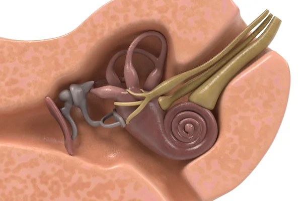
Inner Ear Hair Cells Field In The Vestibular System - Isometric View 3d Illustration
Image, 10.48MB, 10000 × 6600 jpg

Inner Ear Hair Cells In The Vestibular System - Isometric View 3d Illustration
Image, 11.85MB, 10000 × 6600 jpg
Inner Ear Hair Cells In The Vestibular System - Closeup View 3d Illustration
Image, 9.96MB, 10000 × 6600 jpg
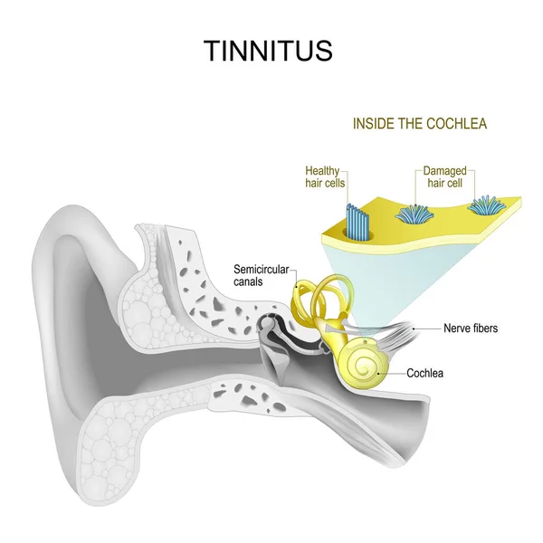
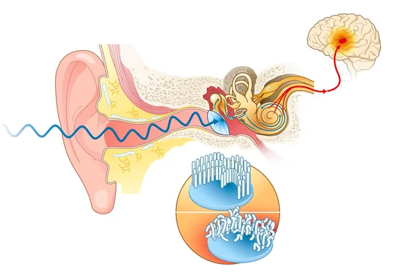
Tinnitus. Human Ear. Part Of Auditory System. Inside The Cochlea. Close-up Of Healthy And Damaged Hair Cells System. Vector Illustration.
Vector, 13.83MB, 4444 × 4443 eps
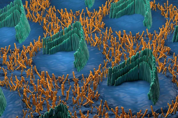
Inner Ear Hair Cells In The Vestibular System - Isometric View 3d Illustration
Image, 10.25MB, 10000 × 6600 jpg
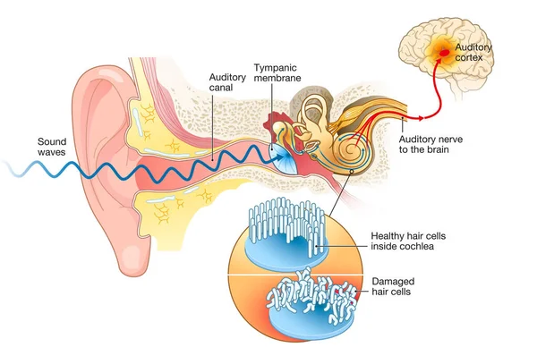
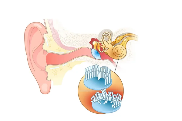
Illustration Showing Tinnitus. Damaged Hair Cells Inside Cochlea, Labeled
Image, 5.82MB, 6837 × 4545 jpg
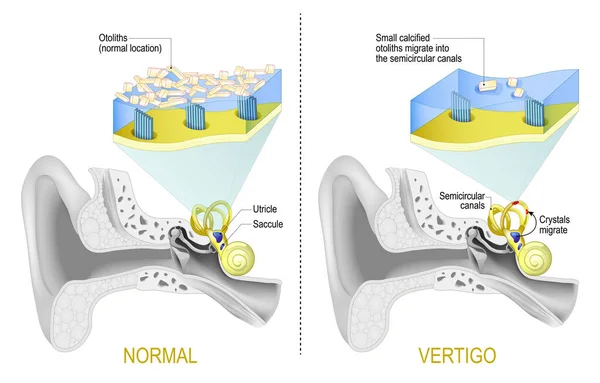
Benign Paroxysmal Positional Vertigo. BPPV Is A Disorder Arising From A Problem In The Inner Ear. Labyrinth Of The Inner Ear With Semicircular Canals. Comparison And Difference Between Normal Vestibular System And Vertigo When Small Calcified Otolith
Vector, 7.33MB, 5000 × 3195 eps

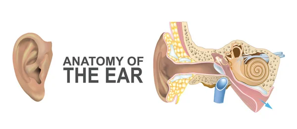
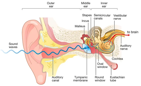
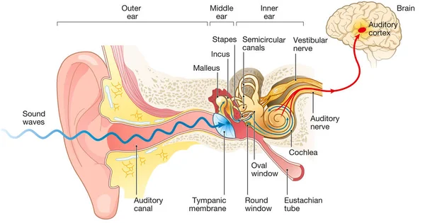
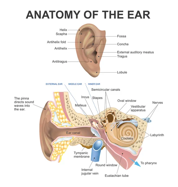
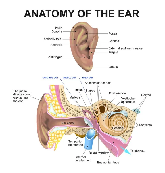
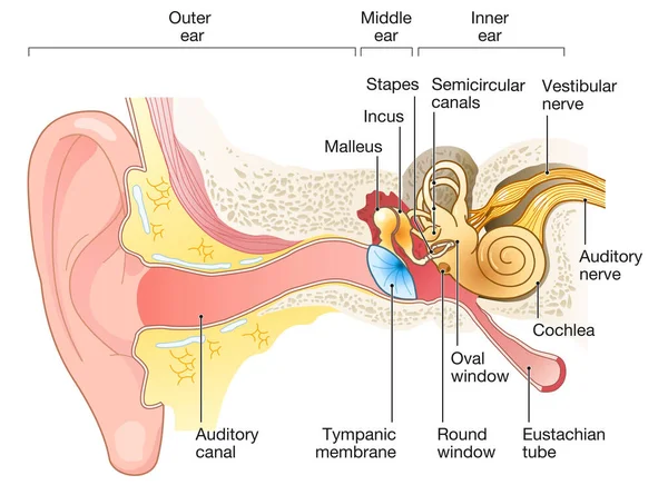
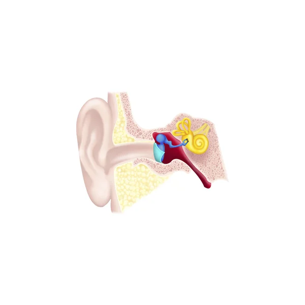
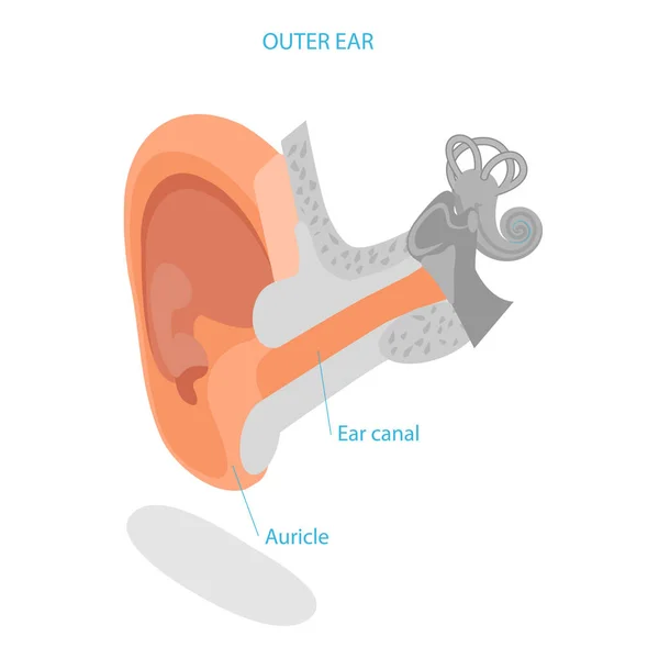
Ear Anatomy. Cross Section Of External (outer), Middle, And Inner Ear Opened. Close-up Of Human Ear Structure. Poster For Education And Medical Use. Vector Illustration. Easy Editable
Vector, 9.09MB, 4444 × 4444 eps

Anatomy Of The Inner Ear. Close-up Of Hair Cells Inside The Cochlea. Vector Poster
Vector, 10.38MB, 4444 × 4444 eps

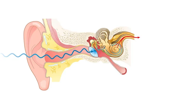
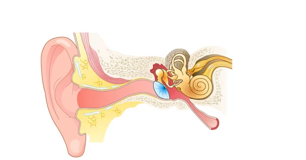
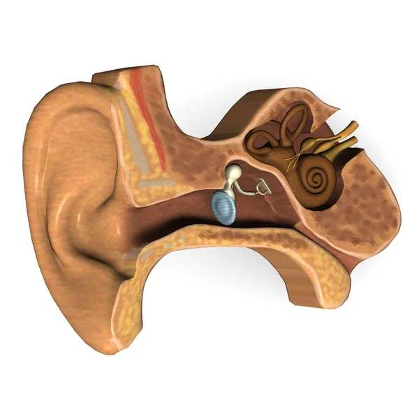
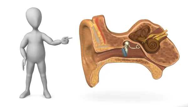
Illustration Showing The Inner Parts Of A Human Ear, Useful For Explaining Hearing Loss Or Auditory System Related Concepts
Vector, 5.44MB, 5000 × 4992 eps
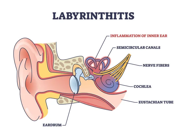
Labyrinthitis As Inner Ear Infection And Medical Inflammation Outline Diagram. Labeled Educational Scheme With Painful Condition And Medical Cause For Hearing And Balance Loss Vector Illustration.
Vector, 6.2MB, 5000 × 3611 eps
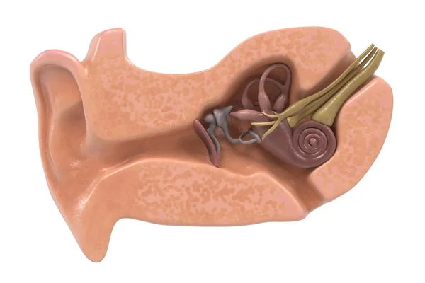
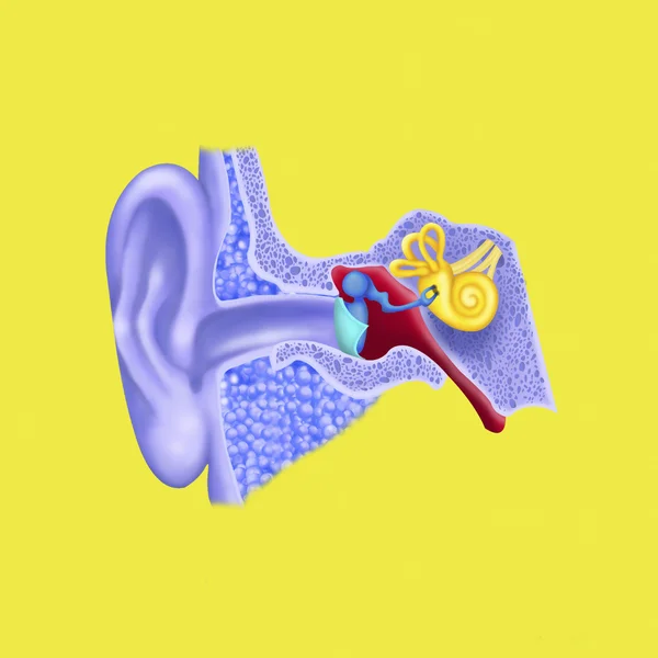
3D Isometric Flat Vector Illustration Of Human Ear Anatomy, Labeled Medical Scheme. Item 3
Vector, 0.67MB, 5000 × 5000 eps
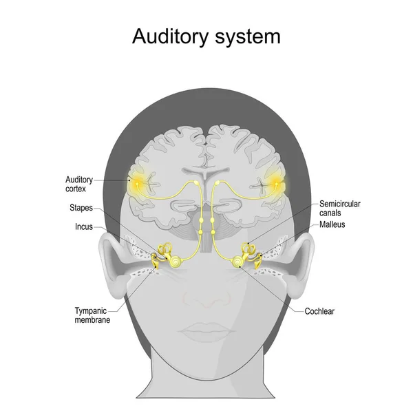
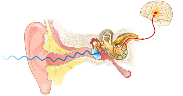
Auditory System From Tympanic Membrane And Cochlear In The Ear To Auditory Cortex On The Brain. Sensory System For The Sense Of Hearing. Anatomy Of The Human Ear. Vector Poster
Vector, 10.78MB, 4444 × 4444 eps
Page 1 >> Next