
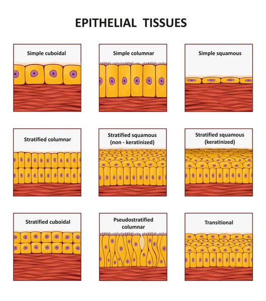
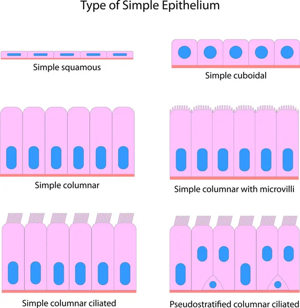
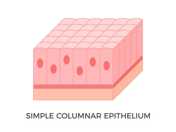
Stock image Simple Epithelium
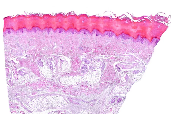
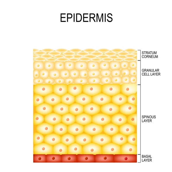
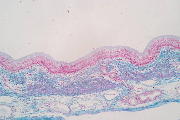
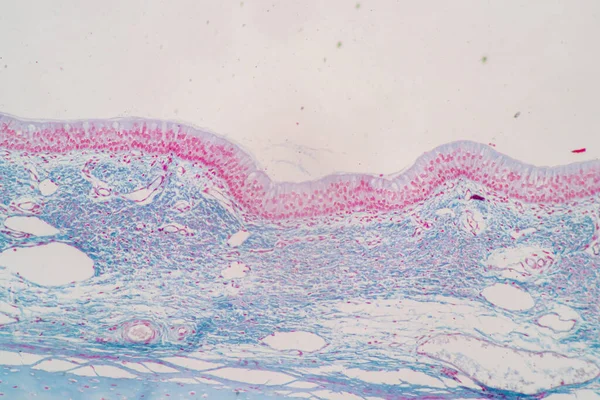
Low Power Light Microscope Micrograph Of Human Glabrous Skin. From Top, The Epidermis, A Keratinized Stratified Squamous Epithelium Showing A Prominent Horny Layer, The Dermis And Hypodermis. The Hypodermis Is Identified By The Presence Of Adipose Ti
Image, 16.01MB, 4587 × 3072 jpg

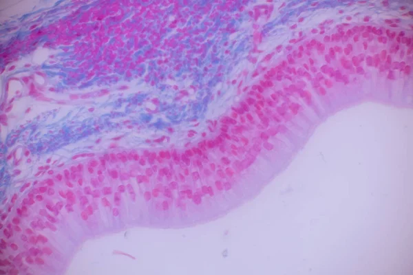
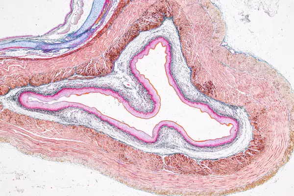
Ciliated Columnar Epithelium. Epithelial Cells Forms The Lining Of The Stomach And Intestines, Duodenum, Fallopian Tubes, Uterus, Central Canal Of The Spinal Cord, Nose, Ears And The Taste Buds.
Vector, 0.98MB, 4444 × 4444 eps
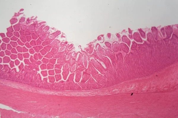
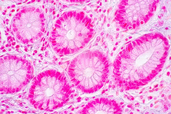
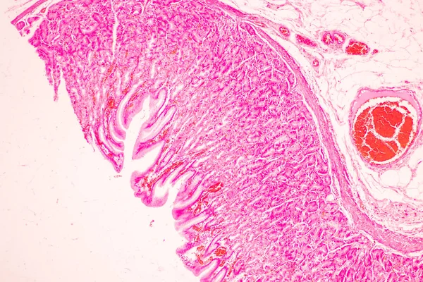
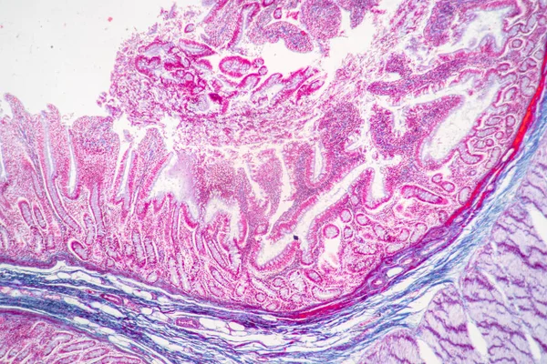
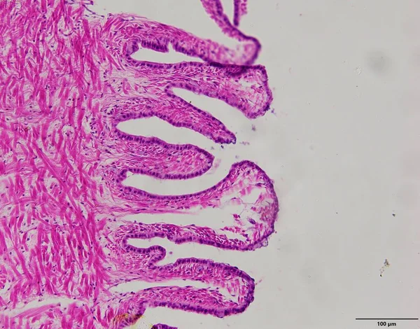
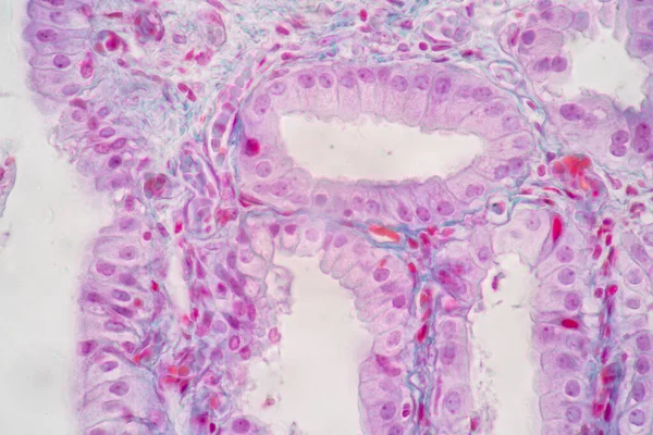
Tissue Of Small Intestine (Duodenum) And Vermiform Appendix Human Under The Microscope In Lab.
Image, 21.54MB, 6000 × 4000 jpg
Tissue Of Small Intestine (Duodenum) And Vermiform Appendix Human Under The Microscope In Lab.
Image, 18.99MB, 6000 × 4000 jpg
Tissue Of Small Intestine (Duodenum) And Vermiform Appendix Human Under The Microscope In Lab.
Image, 22.69MB, 6000 × 4000 jpg

Tissue Of Small Intestine (Duodenum) And Vermiform Appendix Human Under The Microscope In Lab.
Image, 20.31MB, 6000 × 4000 jpg
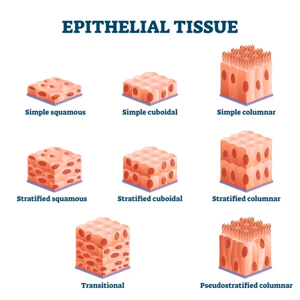
Epithelial Tissue With Labeled Squamous, Cuboidal And Columnar Examples.
Vector, 8.25MB, 4000 × 4000 eps


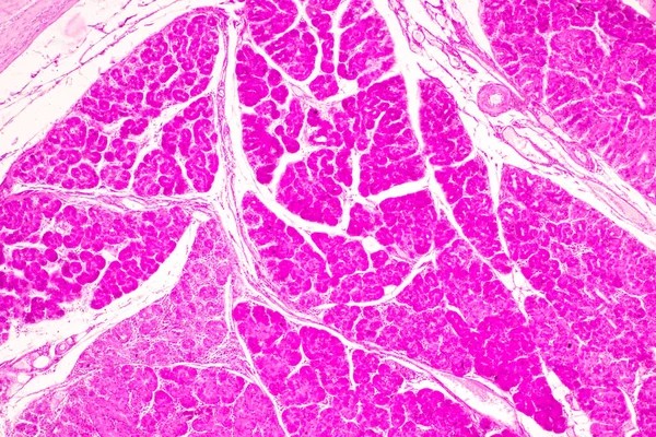
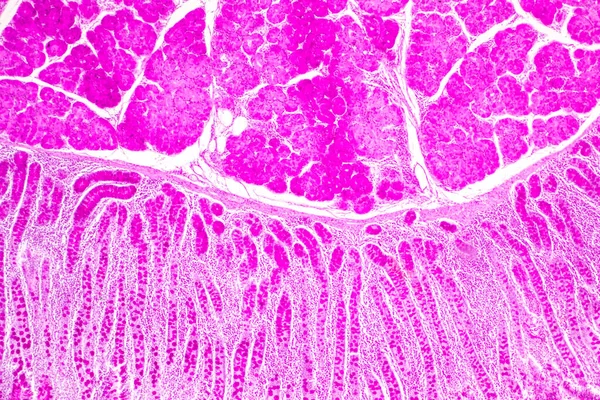
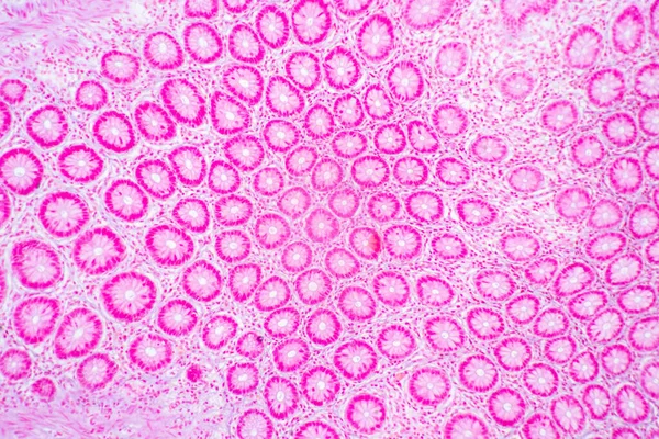
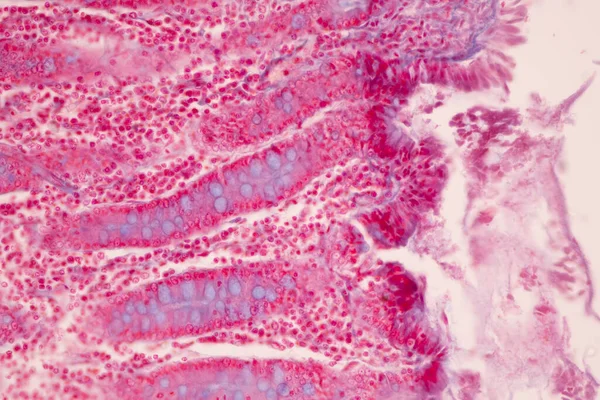
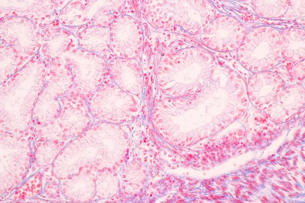
Backgrounds Of Characteristics Tissue Of Stomach Human, Small Intestine Human, Pancreas Human And Large Intestine Human Under The Microscope In Lab.
Image, 20.75MB, 6720 × 4480 jpg

Backgrounds Of Characteristics Tissue Of Stomach Human, Small Intestine Human, Pancreas Human And Large Intestine Human Under The Microscope In Lab.
Image, 20.92MB, 6720 × 4480 jpg
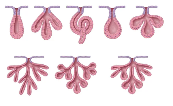
Exocrine Glands Have Two Structural Classifications, Unicellular (one Cell Layer) And Multicellular (many Cell Layers)
Image, 10.06MB, 9449 × 5809 jpg
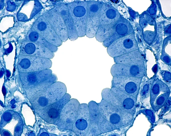
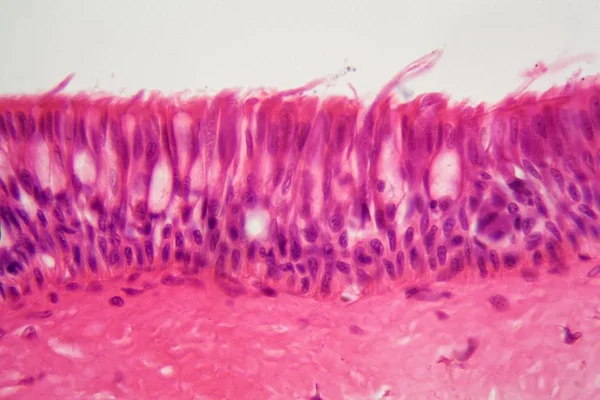
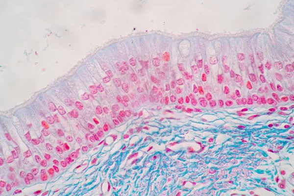
Characteristics Of Columnar Epithellum Cell (Cell Structure) Of Human Under Microscope View For Education In Laboratory.
Image, 16.28MB, 6720 × 4480 jpg


Tissue Of Small Intestine (Duodenum) And Vermiform Appendix Human Under The Microscope In Lab.
Image, 17.16MB, 6000 × 4000 jpg
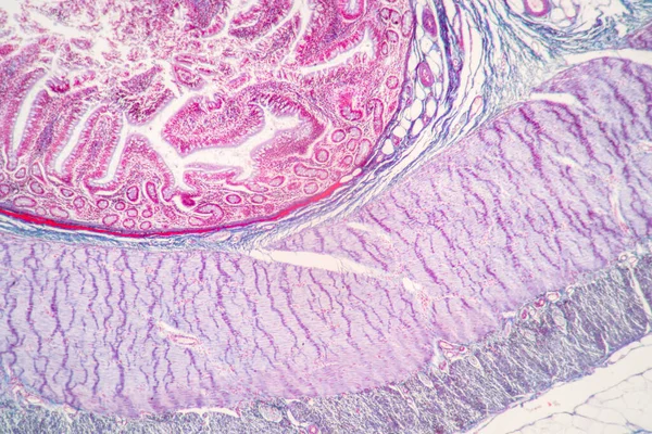
Tissue Of Small Intestine (Duodenum), Large Intestine Human And Stomach Human Under The Microscope In Lab.
Image, 17.29MB, 8192 × 5461 jpg
Backgrounds Of Characteristics Tissue Of Stomach Human, Small Intestine Human, Pancreas Human And Large Intestine Human Under The Microscope In Lab.
Image, 19.31MB, 6720 × 4480 jpg
Characteristics Of Columnar Epithellum Cell (Cell Structure) Of Human Under Microscope View For Education In Laboratory.
Image, 17.03MB, 6720 × 4480 jpg
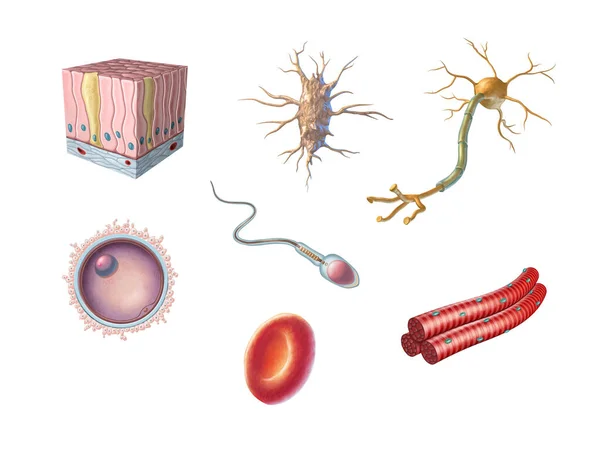
Different Types Of Human Cells Including An Egg Cell, Sperm, Red Blood Cell, Osteocyte, Neuron, Skeletal Muscle And Columnar Epithelial Cell. Digital Illustration.
Image, 5.96MB, 8000 × 6055 jpg
Backgrounds Of Characteristics Tissue Of Stomach Human, Small Intestine Human, Pancreas Human And Large Intestine Human Under The Microscope In Lab.
Image, 20.16MB, 6720 × 4480 jpg

Characteristics Of Columnar Epithellum Cell (Cell Structure) Of Human Under Microscope View For Education In Laboratory.
Image, 17.52MB, 6720 × 4480 jpg

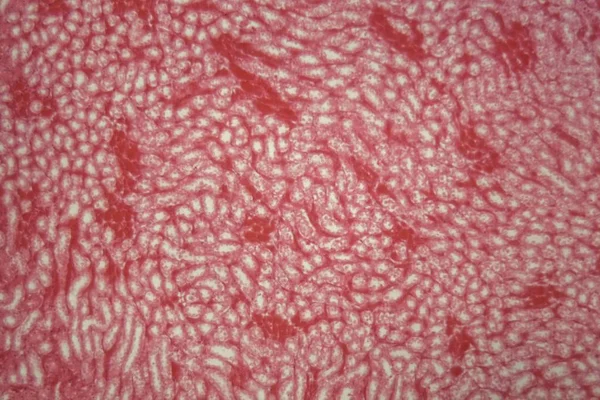
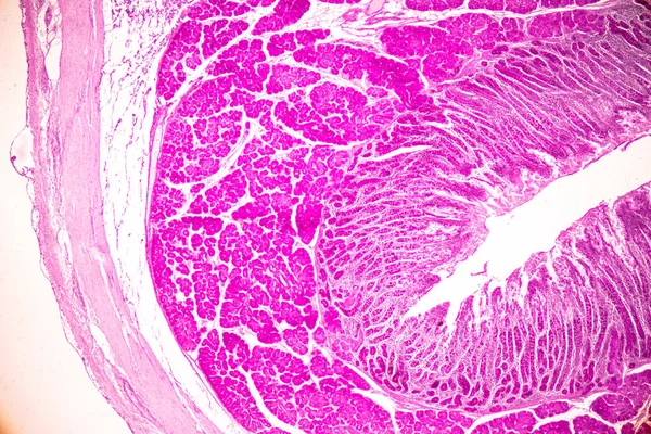
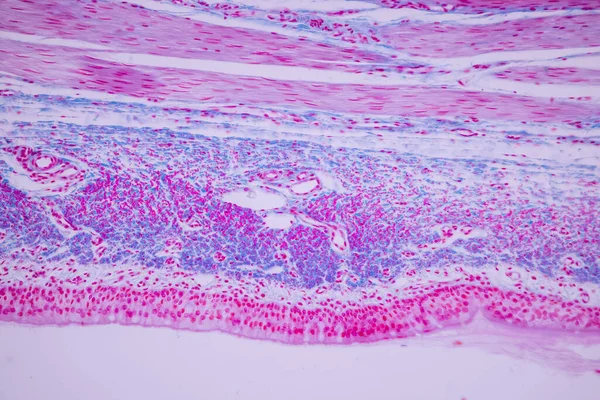
Pathology And Histology Tissue Of Mouse, Rabbit, Cat And Cow Under Microscope.
Image, 32.75MB, 6000 × 4000 jpg

Pathology And Histology Tissue Of Mouse, Rabbit, Cat And Cow Under Microscope.
Image, 6.85MB, 2667 × 4000 jpg
Pathology And Histology Tissue Of Mouse, Rabbit, Cat And Cow Under Microscope.
Image, 18.37MB, 6000 × 4000 jpg

Pathology And Histology Tissue Of Mouse, Rabbit, Cat And Cow Under Microscope.
Image, 8.03MB, 6000 × 3245 jpg

Vector Design Of Biology And Medical Sign. Collection Of Biology And Skeleton Stock Vector Illustration.
Vector, 3.6MB, 5000 × 5000 eps

Characteristics Of Columnar Epithellum Cell (Cell Structure) Of Human Under Microscope View For Education In Laboratory.
Image, 16.38MB, 6720 × 4480 jpg
Simple Columnar Epithelium. Epithelial Tissue Types. Tall And Slender Cells With Oval-shaped Nuclei. Lines Most Organs Of The Digestive Tract Like Stomach, Intestines. Medical Illustration. Vector.
Vector, 4.96MB, 5000 × 3750 eps

Pathology And Histology Tissue Of Mouse, Rabbit, Cat And Cow Under Microscope.
Image, 34.2MB, 6000 × 4000 jpg

Pathology And Histology Tissue Of Mouse, Rabbit, Cat And Cow Under Microscope.
Image, 18.99MB, 6000 × 4000 jpg

Pathology And Histology Tissue Of Mouse, Rabbit, Cat And Cow Under Microscope.
Image, 34.94MB, 6000 × 4000 jpg

Pathology And Histology Tissue Of Mouse, Rabbit, Cat And Cow Under Microscope.
Image, 10.76MB, 6000 × 4000 jpg

Pathology And Histology Tissue Of Mouse, Rabbit, Cat And Cow Under Microscope.
Image, 16.84MB, 6000 × 4000 jpg
Characteristics Of Columnar Epithellum Cell (Cell Structure) Of Human Under Microscope View For Education In Laboratory.
Image, 17.31MB, 6720 × 4480 jpg

Pathology And Histology Tissue Of Mouse, Rabbit, Cat And Cow Under Microscope.
Image, 20.89MB, 6000 × 4000 jpg
Page 1 >> Next