
Stock image Spinal Fusion

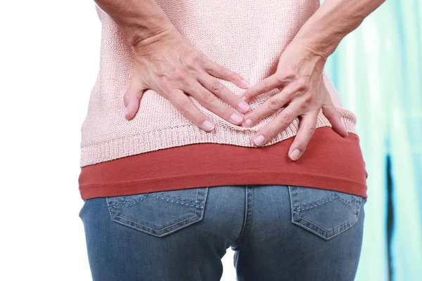
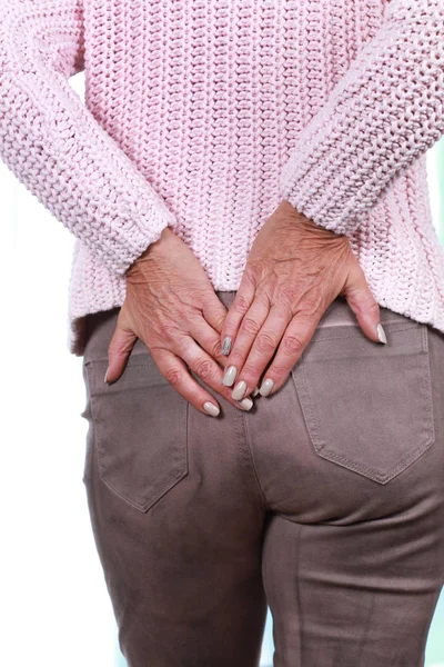
Doctor And Patient Talking About Back Pain Treatment In Clinic Office
Image, 12.16MB, 5748 × 3833 jpg
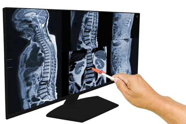
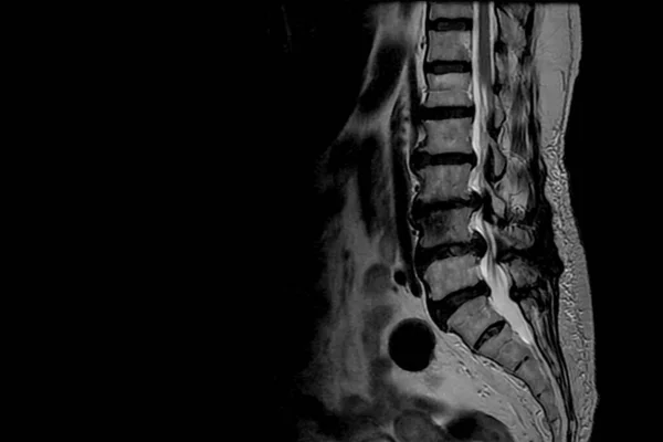
Collection MRI Of Lumbar Spine History Of Fall With Back Pain, Radiate To Leg, Rule Out Spinal Stenosis .Impression:Burst Fracture Of L2 Vertebral Body With Severe Vertebral Collapse.Medical Concept.
Image, 8.31MB, 5820 × 3890 jpg
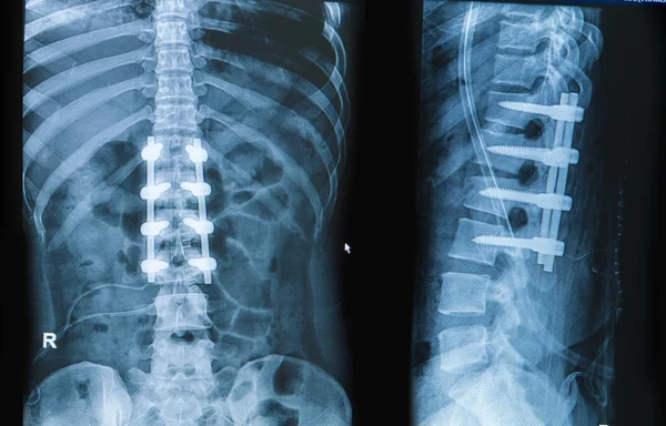
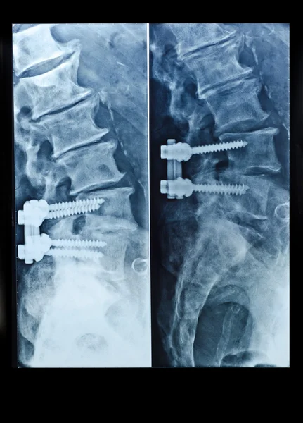
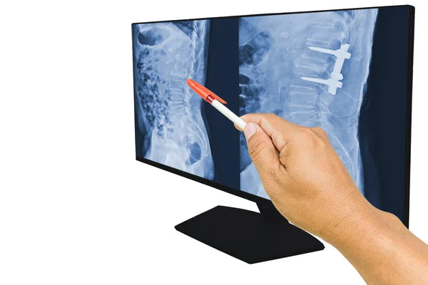
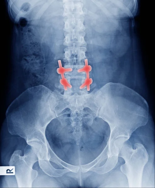
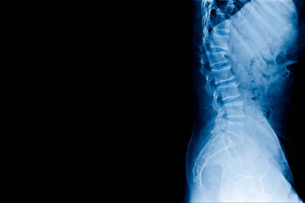
Soft And Blurry X-ray LS-spine Post Fixed Compression Fracture L1 Vertebral Body With Rods. Normal Alignment And Disc Space.
Image, 4.54MB, 6000 × 3317 jpg


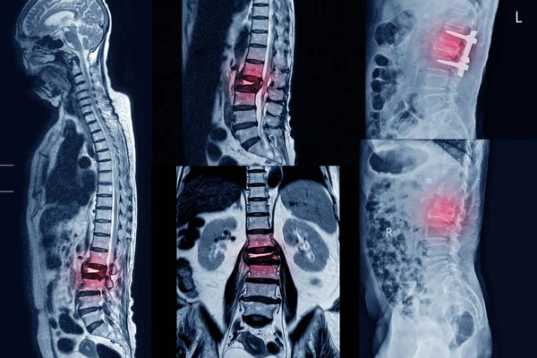
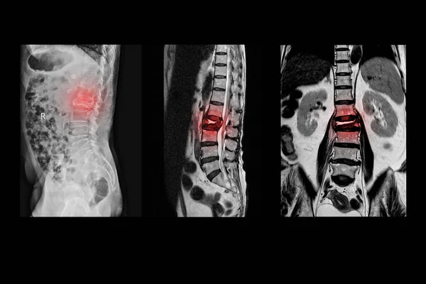
Medical X-ray And MRI Of Lumbar Spine Compression Fracture Bulging Of L1-2. On Arrow Point..Lumbar Spondylosis From L1-2 To L5-S1 Discs.Medical Healthcare Concept.
Image, 6.06MB, 5500 × 3500 jpg
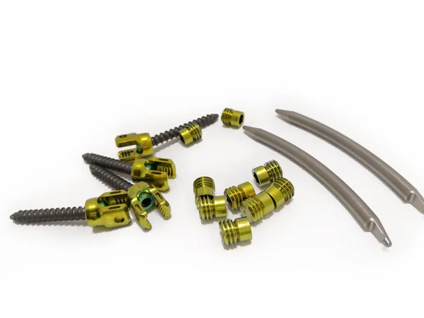
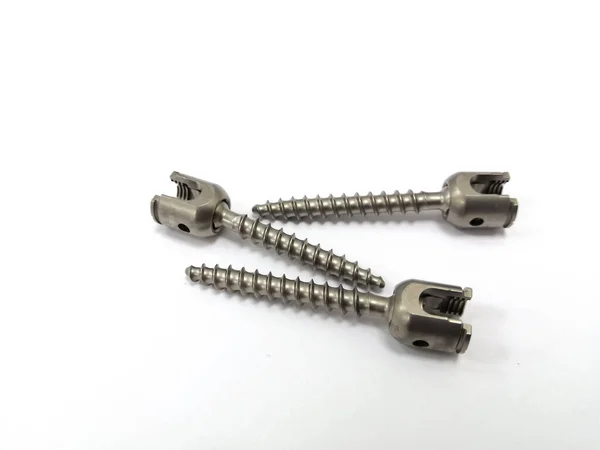
Pedicle Screws, Nuts And Rods In Isolated Background. Using For Spinal Fusion Surgery
Image, 3.14MB, 4500 × 3375 jpg

MRI Of Lumbar Spine The Study Reveals Burst Fracture Of L2 Vertebral Body, Appears As Severe Decreased Disc Height And Widening Of Interpedicular Distance.
Image, 2.77MB, 3096 × 4128 jpg
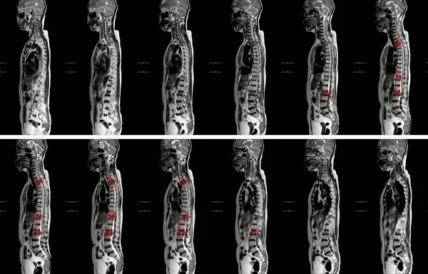
MRI OF THORACOLUMBAR SPINE IMPRESSION: Moderate Pathological Compression Of T11 And L2 Levels With Enhancing Multiple Marrow Lesions At T1, T10 ToT12, L2, L3 To L5 Levels.
Image, 4.96MB, 5688 × 3643 jpg
Cardiologist Doctor Holding Stethoscopet, Blurred Electrocardiogram Result On Paper With AICD Pacemaker In Chest X-ray Background.medical Concept
Image, 5.46MB, 5500 × 3500 jpg

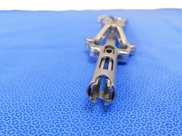
Closeup Image Of Spinal Surgical Instrument Beale Rod Reducer Short. Selective Focus
Image, 6.9MB, 4500 × 3375 jpg

MRI Of Lumbar Spine History Of Fall With Back Pain, Radiate To Leg, Rule Out Spinal Stenosis .Burst Fracture Of L2 Vertebral Body With Severe Vertebral Collapse.
Image, 4.67MB, 6000 × 4000 jpg

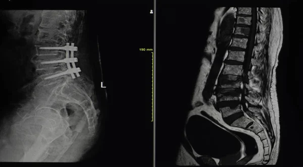
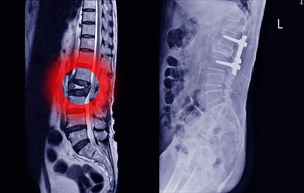
The Doctor Reported The MRI Scans Of The Lumbar Spine Compression Fracture Bulging Of L1-L2. And Post Operation Fixed By Iron Rod And Screws. Medical Education Concept.
Image, 6.18MB, 6000 × 3792 jpg
The Doctor Reported The MRI Scans Of The Lumbar Spine Compression Fracture Bulging Of L1-L2. And Post Operation Fixed By Iron Rod And Screws. Medical Education Concept.
Image, 7.51MB, 6000 × 4000 jpg
The Doctor Reported The MRI Scans Of The Lumbar Spine Compression Fracture Bulging Of L1-L2. And Post Operation Fixed By Iron Rod And Screws. Medical Education Concept.
Image, 6.16MB, 6000 × 4000 jpg
Closeup Image Of Thoracic Lumbar Vertebra Pedicle Screw In White Background
Image, 2.69MB, 4200 × 3150 jpg

Spine Posterior Thoracic Fusion With Pedicle Screws And Rods On Blue Background
Image, 3.12MB, 6000 × 3600 jpg
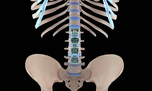
3D Medical Illustration Of Orthopedic Spine Fixation Spinal Fixator Anterior Lumbar Plate
Image, 2.6MB, 5000 × 3000 jpg
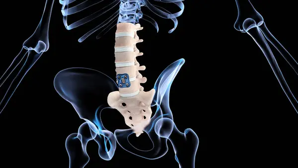
3D Medical Illustration Of Orthopedic Spine Fixation Spinal Fixator Anterior Lumbar Plate
Image, 3.49MB, 6000 × 3400 jpg
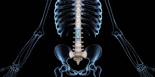
3D Medical Illustration Of Orthopedic Spine Fixation Spinal Fixator Anterior Lumbar Plate
Image, 3.39MB, 6000 × 3000 jpg

3D Medical Illustration Of Orthopedic Spine Fixation Spinal Fixator Anterior Lumbar Plate
Image, 5.33MB, 7000 × 3950 jpg
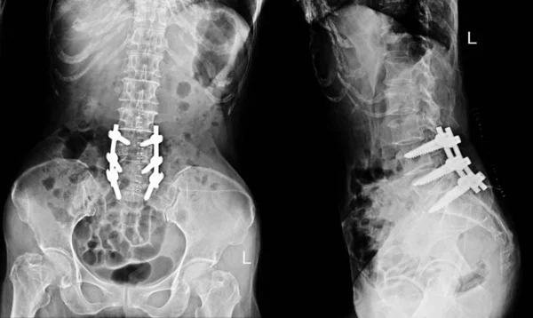
X-ray Lower Back Of Young Man Post Fixed Of Lumbar Spine Fracture, Compression Fracture Of L4-5 Spine With Post Operation
Image, 2.05MB, 1996 × 2428 jpg
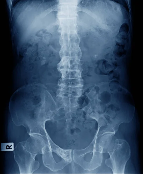
Scan Of Lumbosacral Spine In Lateral Projection Shown Spinal Canal Stenosis .Decrease In Disc Space , Bony Spur Formation And Blank Area At Right Side. Spinal Cord Compression.
Image, 0.28MB, 3000 × 2000 jpg
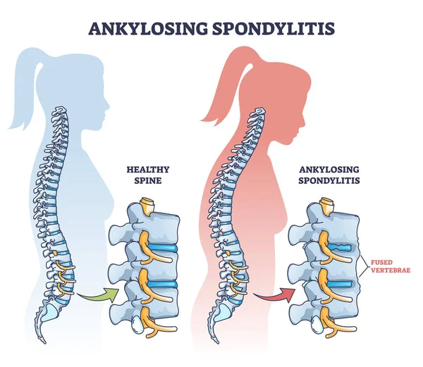
Ankylosing Spondylitis As Inflammatory Spine Bone Disease Outline Diagram. Labeled Educational Anatomical Comparison With Healthy And Damaged Vertebrae Vector Illustration. Fused Skeletal Back Parts.
Vector, 9.4MB, 4600 × 4000 eps
Orthopedic Surgery Related, Pixel Perfect, Editable Stroke, Up Scalable Square Line Vector Icon Set.
Vector, 0.99MB, 7917 × 5000 eps
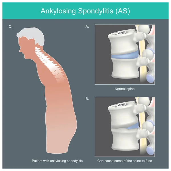
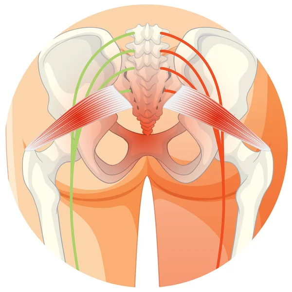
Ankylosing Spondylitis. Human Spine Deformities From Inflammation And Can Cause Some Of The Spine To Fuse
Vector, 1.31MB, 5000 × 5000 ai

Anterior View Of Structure Bone With Internal Fixation For Prevent Compression Spine Bone
Image, 1.89MB, 1996 × 2428 jpg
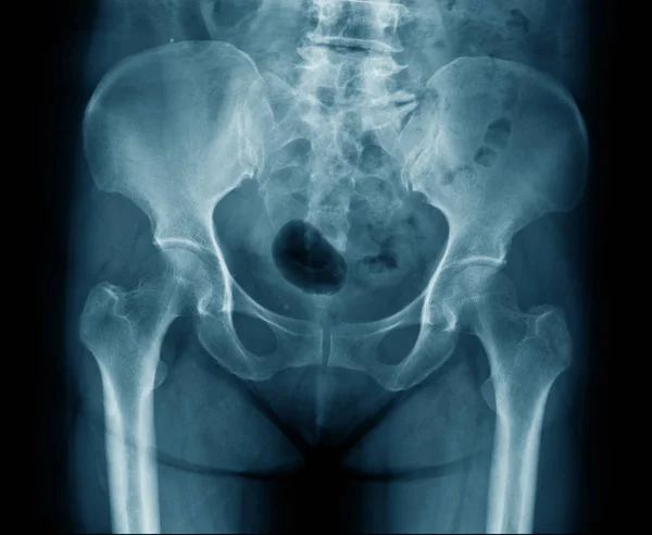
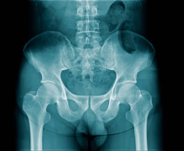
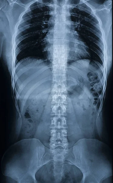
X-ray Image Of Old Man Show Ankylosing Spondylitis Or Bamboo Spine And Degenerative Change Of Human Spine And Pelvic Bone
Image, 1.69MB, 1996 × 2428 jpg

Page 1 >> Next