
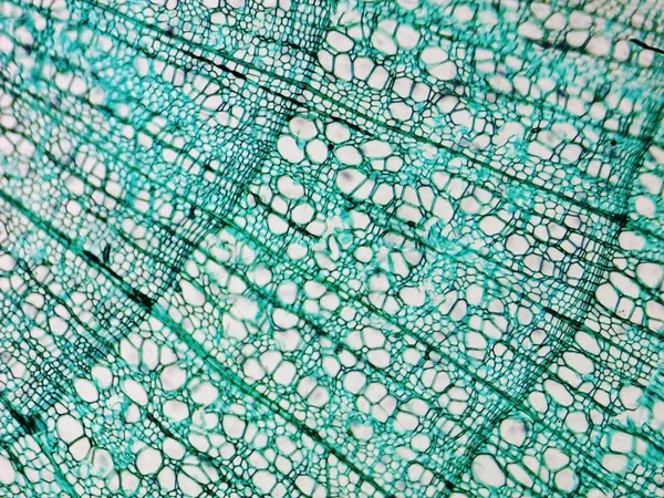
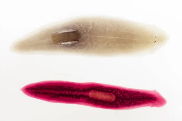
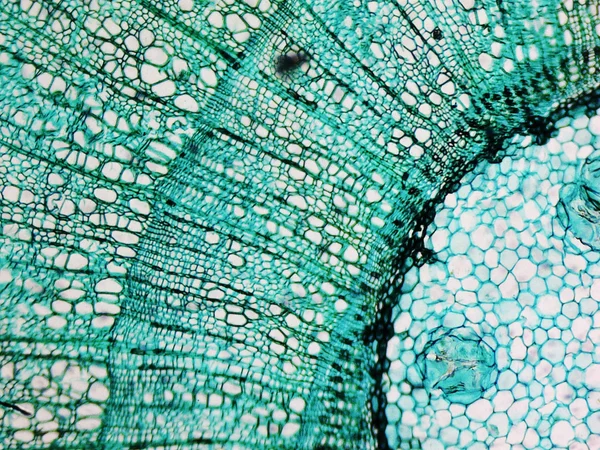
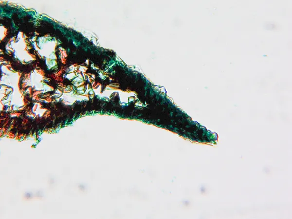
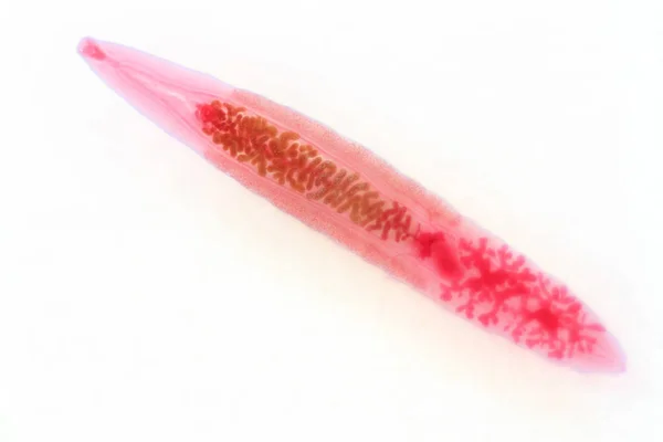
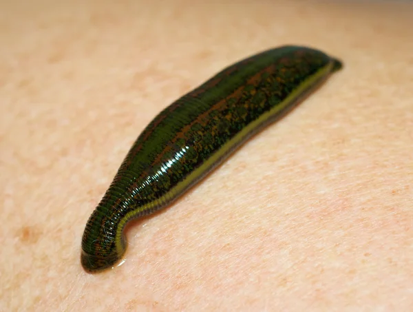
Stock image Stained Slide page 2


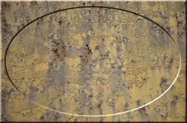
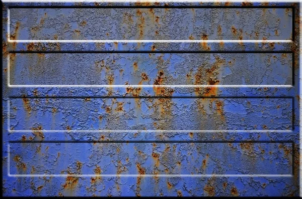
Texture Of Yellow Rough Rusted Metal Surface With Bulky Gray Highlighted Portions Which Can Be Seen On Exposure To Light. Preparation For The Background Processing Of Slides And Spreadsheets
Image, 3.94MB, 2800 × 1855 jpg
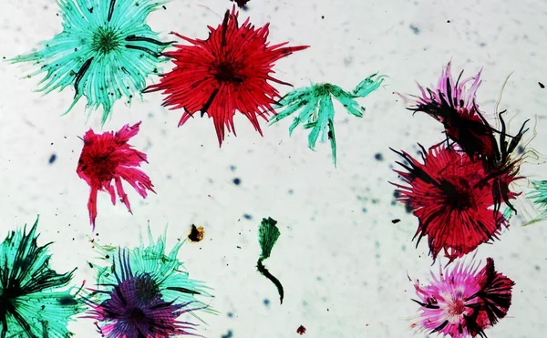
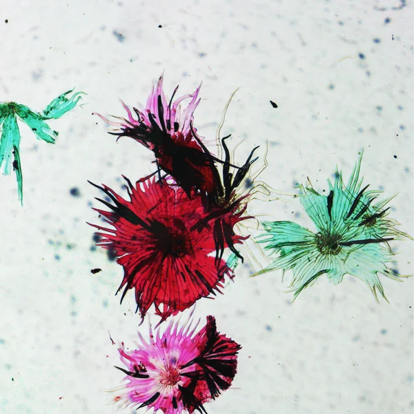


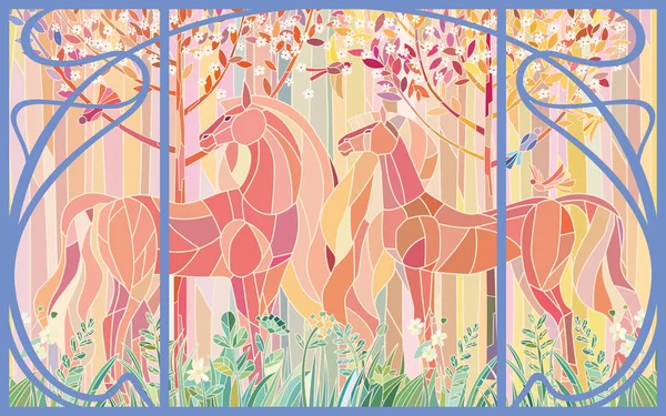
Stained Glass Horses Of Color Patches In The Frame Of Art Nouveau Style. Delicate Shades Of Pink Orange Green. Imitation Colored Glass
Vector, 5.75MB, 6000 × 3750 eps
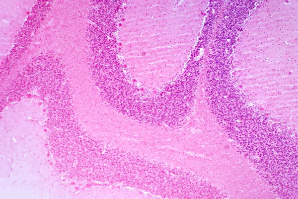
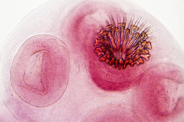
Cerebellum Cross Section Tissue Under The Light Microscope For Pathology Education. Haematoxylin And Eosin Staining Technique For Human Tissue.
Image, 22.42MB, 8192 × 5464 jpg

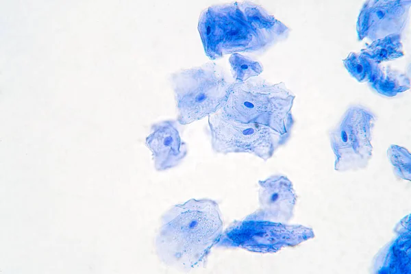
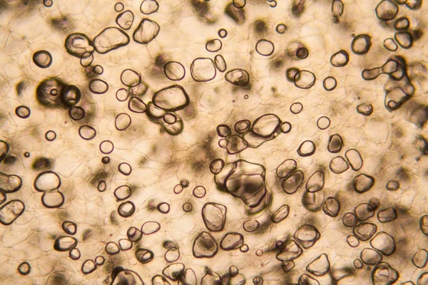
Human Cheek Epithelial Cells. The Tissue That Lines The Inside Of The Mouth Is Known As The Basal Mucosa And Is Composed Of Squamous Epithelial Cells. Education Pathology.
Image, 10.51MB, 6000 × 4000 jpg

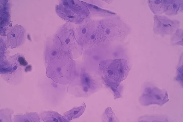
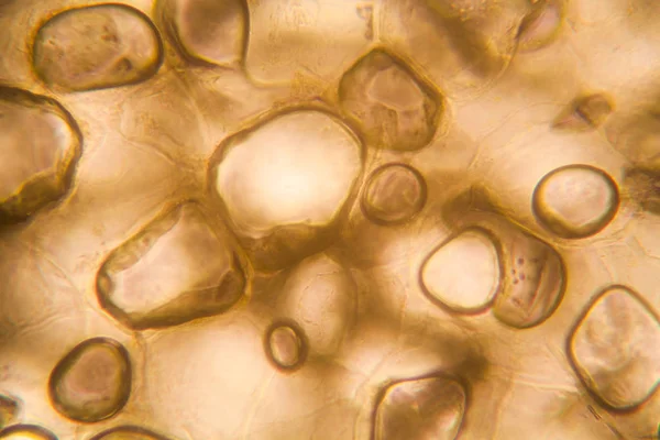
Human Cheek Epithelial Cells. The Tissue That Lines The Inside Of The Mouth Is Known As The Basal Mucosa And Is Composed Of Squamous Epithelial Cells. Education Pathology.
Image, 16.77MB, 6000 × 4000 jpg


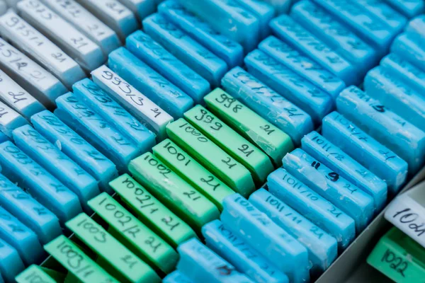
Scientist Is Preparing A Tumor Slide. Microscopy Of Cytopathology Slides And Pathology.
Image, 12.02MB, 6000 × 4000 jpg


Light Photomicrograph Of An Onion Epidermus Cells Seen Through A Microscope
Image, 1.06MB, 2900 × 2172 jpg

Previous << Page 2 >> Next