photo free
Home Images Vectors Videos

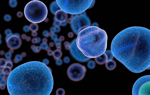
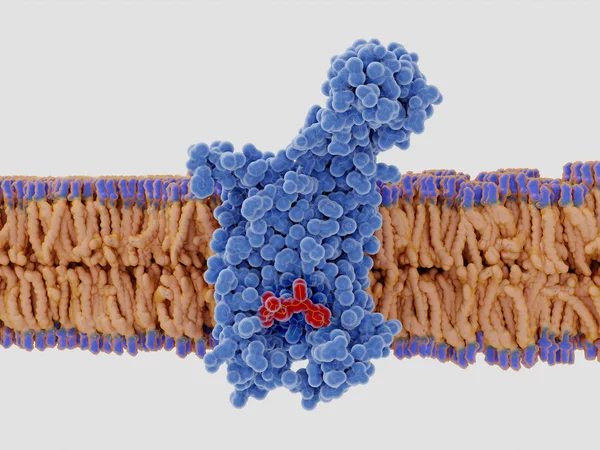
Stock image T Cell Receptor

stock vector CAR T cell therapy. cancer immunotherapy. Artificial leukocyte r

CAR T Cell Therapy. Cancer Immunotherapy. Artificial Leukocyte R

Vector, 6.7MB, 4021 × 4021 eps
stock vector Activation of  T-cell leukocytes

Activation Of T-cell Leukocytes

Vector, 9.93MB, 4992 × 4992 eps
stock image T-lymphocytes and dendritic cells, 3D-rendering; Dendritic cells are antigen-presenting cells of the immune system. They process antigen material and present it on the cell surface to the T-cells. Illustration

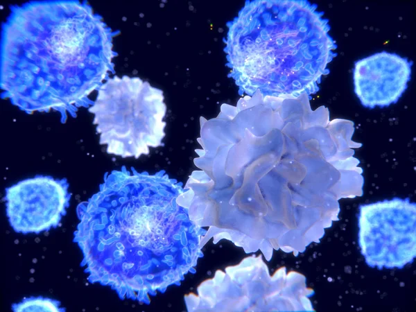
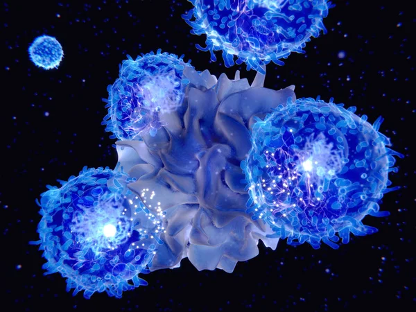
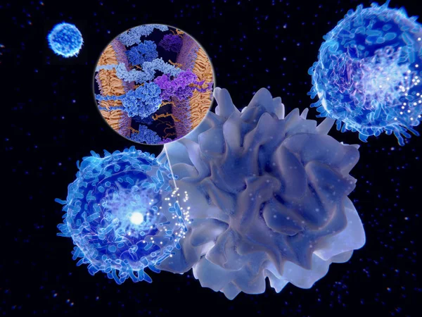
T-lymphocytes And Dendritic Cells, 3D-rendering; Dendritic Cells Are Antigen-presenting Cells Of The Immune System. They Process Antigen Material And Present It On The Cell Surface To The T-cells. Illustration

Image, 2.23MB, 4000 × 3000 jpg
stock vector CAR - Chimeric antigen receptor T cell. lymphocyte that have been genetically engineered to produce an artificial T-cell receptor for use in immunotherapy. Treatment of cancer. Vector illustration

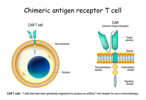
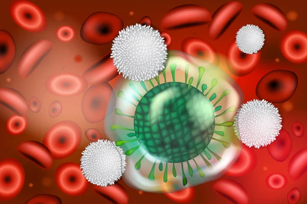
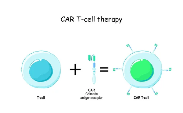
CAR - Chimeric Antigen Receptor T Cell. Lymphocyte That Have Been Genetically Engineered To Produce An Artificial T-cell Receptor For Use In Immunotherapy. Treatment Of Cancer. Vector Illustration

Vector, 6.18MB, 5000 × 3443 eps
stock vector B-cell and T helper cells function.

B-cell And T Helper Cells Function.

Vector, 12.36MB, 5524 × 5524 eps
stock image Engineered receptors (light blue) on the surface of a T-lymphocyte bind specifically to CD19-antigen molecules (magenta) on a leukemia cell. This activates a signal cascade in the T-cell leading to the segregation of vesicles that contain perforin an

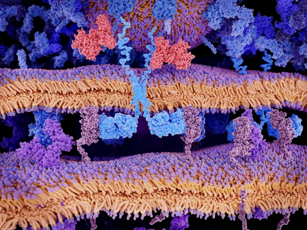
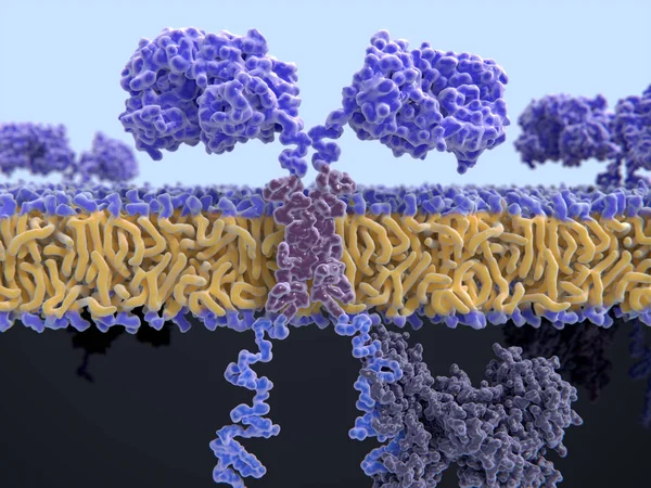
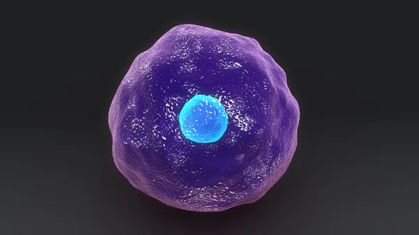
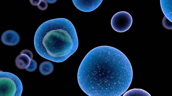
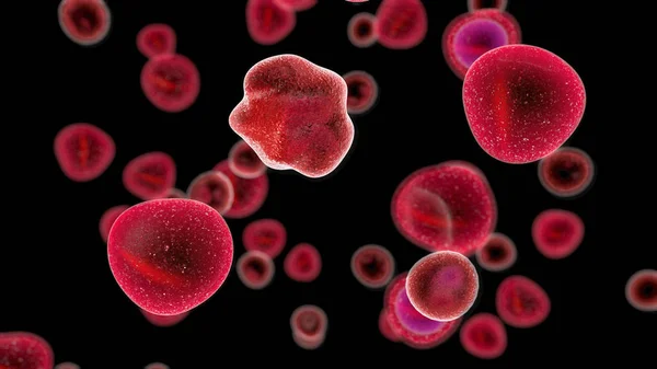
Engineered Receptors (light Blue) On The Surface Of A T-lymphocyte Bind Specifically To CD19-antigen Molecules (magenta) On A Leukemia Cell. This Activates A Signal Cascade In The T-cell Leading To The Segregation Of Vesicles That Contain Perforin An

Image, 11.44MB, 8000 × 6000 jpg
stock image Human Cytotoxic T cell

Human Cytotoxic T Cell

Image, 5.72MB, 3840 × 2160 jpg
stock image T Cells attacking Cancer Cells, 3D illustration

T Cells Attacking Cancer Cells, 3D Illustration

Image, 12.73MB, 4773 × 2770 jpg
stock vector T-cell and B-cell. Cells of Adaptive immune system. immune response and lymphocytes. Vector illustration on white background.

T-cell And B-cell. Cells Of Adaptive Immune System. Immune Response And Lymphocytes. Vector Illustration On White Background.

Vector, 0.75MB, 5000 × 3671 eps
stock image Human Cytotoxic T cell

Human Cytotoxic T Cell

Image, 3.3MB, 3840 × 2160 jpg
stock image T Cells attacking Cancer Cells 3D illustration

T Cells Attacking Cancer Cells 3D Illustration

Image, 4.33MB, 3546 × 2160 jpg
stock image T Cells attacking Cancer Cells, 3D illustration

T Cells Attacking Cancer Cells, 3D Illustration

Image, 9.04MB, 4773 × 2770 jpg
stock image T Cells attacking Cancer Cells 3D illustration

T Cells Attacking Cancer Cells 3D Illustration

Image, 4.34MB, 3840 × 2160 jpg
stock image Lymphocyte Cell Concept

Lymphocyte Cell Concept

Image, 4.97MB, 4150 × 4150 jpg
stock image T Cells attacking Cancer Cells 3D illustration

T Cells Attacking Cancer Cells 3D Illustration

Image, 4.41MB, 3840 × 2160 jpg
stock image 3d computer illustration of a chimeric antigen receptor. CARs are engineered cell receptors that allow T cells to recognize and attack cancer cells in a specific way. They are built by connecting several functional parts from different proteins.

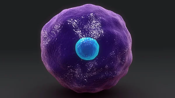
3d Computer Illustration Of A Chimeric Antigen Receptor. CARs Are Engineered Cell Receptors That Allow T Cells To Recognize And Attack Cancer Cells In A Specific Way. They Are Built By Connecting Several Functional Parts From Different Proteins.

Image, 8.45MB, 8000 × 6000 jpg
stock image Interactions of MHC-II with the T-cell receptor and CD4  and B7-1 with CD-28 activates T-cells while the interactions of P7-1 with CTLA-4  and PD-L1  with PD-1 deactivates T-cells.

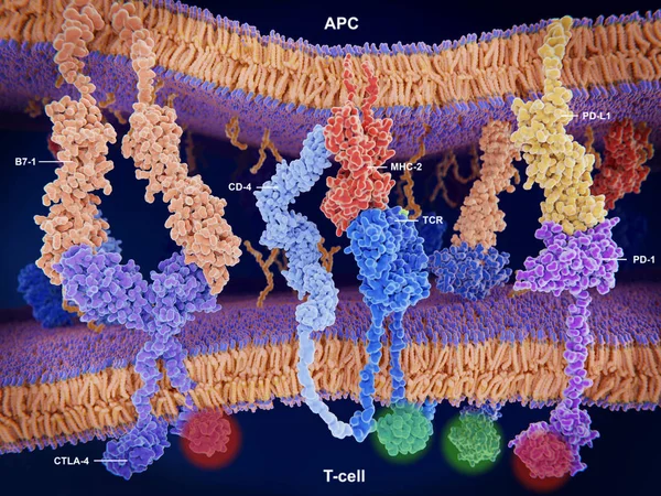
Interactions Of MHC-II With The T-cell Receptor And CD4 And B7-1 With CD-28 Activates T-cells While The Interactions Of P7-1 With CTLA-4 And PD-L1 With PD-1 Deactivates T-cells.

Image, 10.7MB, 8000 × 6000 jpg
stock image Lymphocyte cell concept as an immune system cell representing the control of cancer through immunology or immunotherapy as an oncology medicine symbol on black as a 3D illustration.

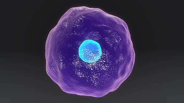
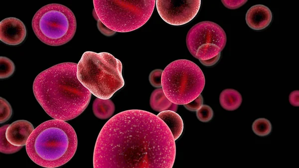
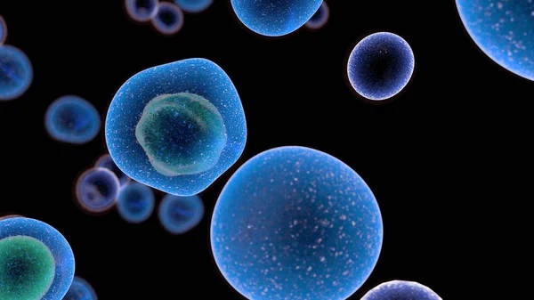
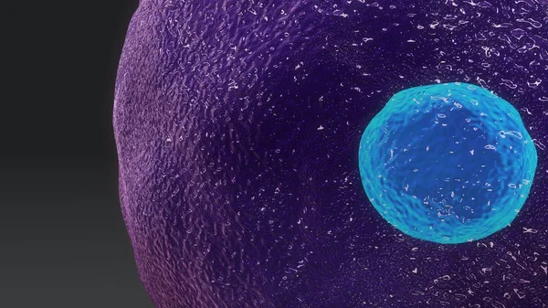
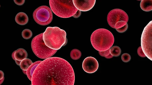
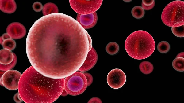
Lymphocyte Cell Concept As An Immune System Cell Representing The Control Of Cancer Through Immunology Or Immunotherapy As An Oncology Medicine Symbol On Black As A 3D Illustration.

Image, 4.93MB, 4232 × 4232 jpg
stock image 3d computer illustration of a dendritic cell. They areantigen-presenting cells  of the immune system. Their main function is to process antigen material and present it on the cell surface to the T cells of the immune system. They are messengers betwe

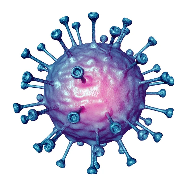
3d Computer Illustration Of A Dendritic Cell. They Areantigen-presenting Cells Of The Immune System. Their Main Function Is To Process Antigen Material And Present It On The Cell Surface To The T Cells Of The Immune System. They Are Messengers Betwe

Image, 5.2MB, 8000 × 6000 jpg
stock vector Lymphocytes attacking viruses. 3D medical illustration on immunity

Lymphocytes Attacking Viruses. 3D Medical Illustration On Immunity

Vector, 8.58MB, 5000 × 3329 eps
stock image Human Cytotoxic T cell

Human Cytotoxic T Cell

Image, 3.37MB, 3840 × 2160 jpg
stock vector Types of Lymphocytes. T and B cell. Human adaptive immune system. immune response and component of humoral immunity. Antibody, Plasma cell, T helper, and T-killer. Vector illustration

Types Of Lymphocytes. T And B Cell. Human Adaptive Immune System. Immune Response And Component Of Humoral Immunity. Antibody, Plasma Cell, T Helper, And T-killer. Vector Illustration

Vector, 1.63MB, 4444 × 4444 eps
stock image Human Cytotoxic T cell

Human Cytotoxic T Cell

Image, 3.35MB, 3840 × 2160 jpg
stock image T Cells attacking Cancer Cells 3D illustration

T Cells Attacking Cancer Cells 3D Illustration

Image, 5.51MB, 3408 × 2160 jpg
stock image T Cells attacking Cancer Cells 3D illustration

T Cells Attacking Cancer Cells 3D Illustration

Image, 4.73MB, 3324 × 2160 jpg
stock image T Cells attacking Cancer Cells 3D illustration

T Cells Attacking Cancer Cells 3D Illustration

Image, 3.8MB, 3840 × 2160 jpg
stock image T Cells attacking Cancer Cells 3D illustration

T Cells Attacking Cancer Cells 3D Illustration

Image, 4.21MB, 3840 × 2160 jpg
stock image T Cells attacking Cancer Cells 3D illustration

T Cells Attacking Cancer Cells 3D Illustration

Image, 3.75MB, 3840 × 2160 jpg
stock image T-cells attacking a cancer cell. Isolated on black background. 3d render

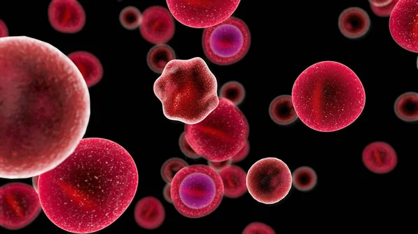
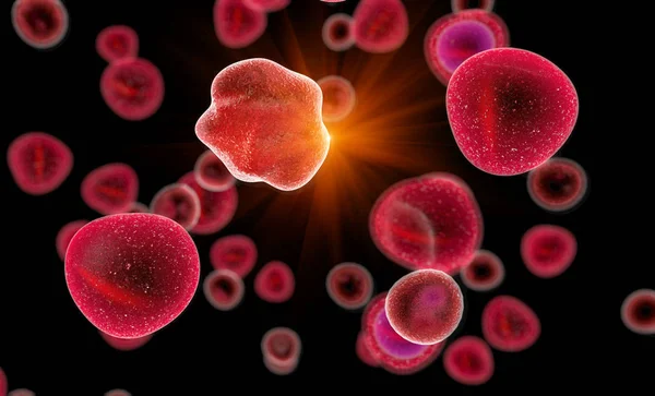
T-cells Attacking A Cancer Cell. Isolated On Black Background. 3d Render

Image, 4.29MB, 4000 × 3000 jpg
stock image T Cells attacking Cancer Cells 3D illustration

T Cells Attacking Cancer Cells 3D Illustration

Image, 4.06MB, 3840 × 2160 jpg
stock image T Cells attacking Cancer Cells 3D illustration

T Cells Attacking Cancer Cells 3D Illustration

Image, 4.2MB, 3840 × 2160 jpg
stock vector Dendritic cells vector illustration. Anatomical labeled closeup scheme with progenitor, immature, nucleus and membrane extensions. Antigen, receptor and T cell diagram.

Dendritic Cells Vector Illustration. Anatomical Labeled Closeup Scheme With Progenitor, Immature, Nucleus And Membrane Extensions. Antigen, Receptor And T Cell Diagram.

Vector, 4.58MB, 4800 × 3386 eps
stock vector T cell and Chimeric antigen receptor (CAR).

T Cell And Chimeric Antigen Receptor (CAR).

Vector, 0.92MB, 5555 × 3441 eps
stock image T Cells attacking Cancer Cells 3D illustration

T Cells Attacking Cancer Cells 3D Illustration

Image, 4.69MB, 3840 × 2160 jpg
stock vector T-cell with Chimeric antigen receptor (CAR t cell) killing of th

T-cell With Chimeric Antigen Receptor (CAR T Cell) Killing Of Th

Vector, 1.14MB, 8582 × 8582 eps
stock vector Innate immunity. Adaptive specific . Phagocytosis. Infographics. vector

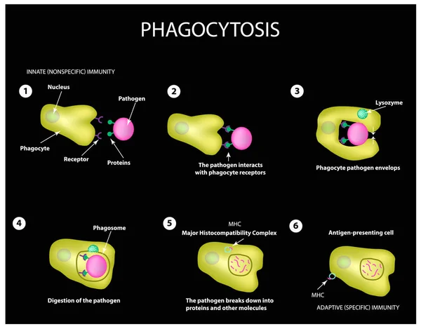
Innate Immunity. Adaptive Specific . Phagocytosis. Infographics. Vector

Vector, 3.99MB, 5000 × 3911 eps
stock image T Cells attacking Cancer Cells 3D illustration

T Cells Attacking Cancer Cells 3D Illustration

Image, 3.9MB, 3840 × 2160 jpg
stock image Human Cytotoxic T cell

Human Cytotoxic T Cell

Image, 5.07MB, 3840 × 2160 jpg
stock image T Cells attacking Cancer Cells 3D illustration

T Cells Attacking Cancer Cells 3D Illustration

Image, 4.06MB, 3564 × 2160 jpg
stock image T Cells attacking Cancer Cells 3D illustration

T Cells Attacking Cancer Cells 3D Illustration

Image, 3.76MB, 3840 × 2160 jpg
stock image T Cells attacking Cancer Cells 3D illustration

T Cells Attacking Cancer Cells 3D Illustration

Image, 4.07MB, 3840 × 2160 jpg
stock vector Cytotoxic cells. Cytokines. Cell immunity. Infographics. Vector illustration

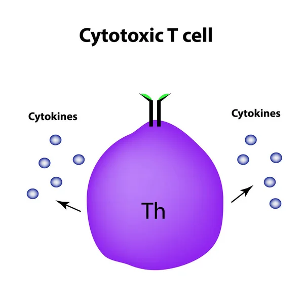
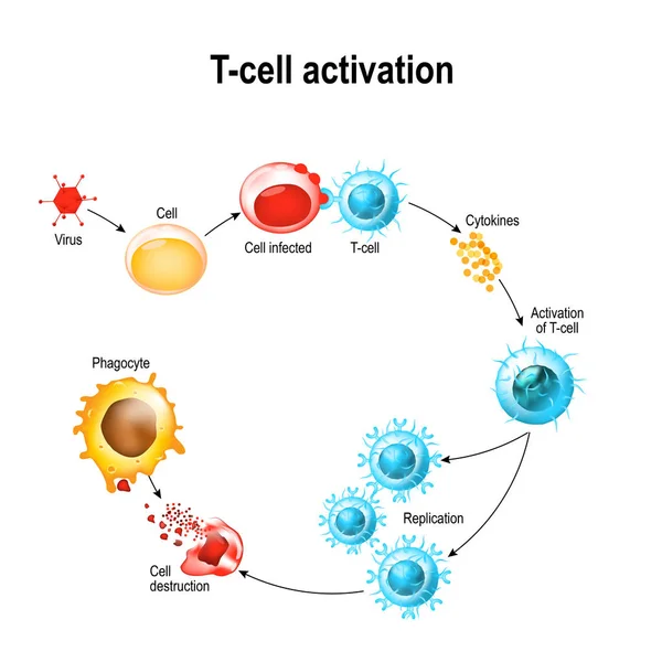
Cytotoxic Cells. Cytokines. Cell Immunity. Infographics. Vector Illustration

Vector, 1.39MB, 5000 × 5000 eps
stock vector T-cell dependent b-cell activation. B lymphocyte binds an antigen, receive help from a T helper, and differentiate into a plasma cell that secretes of antibodies. Receptors on surface of white blood cells. Human immune system. Vector poster

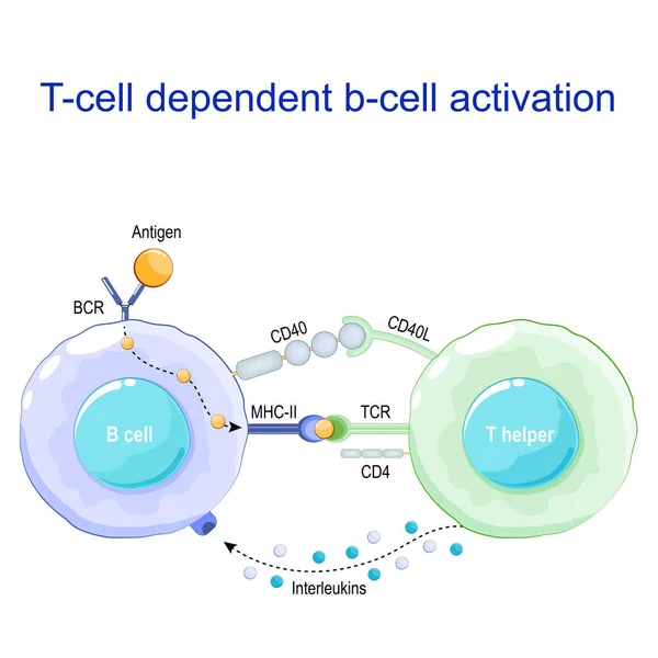
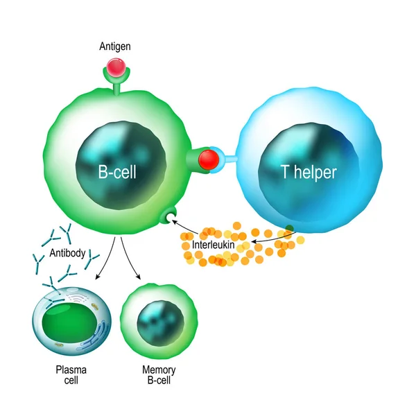
T-cell Dependent B-cell Activation. B Lymphocyte Binds An Antigen, Receive Help From A T Helper, And Differentiate Into A Plasma Cell That Secretes Of Antibodies. Receptors On Surface Of White Blood Cells. Human Immune System. Vector Poster

Vector, 1.05MB, 4444 × 4444 eps
stock vector The life cycle of HIV. Infographics. World AIDS Day. Vector illustration

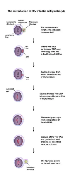
The Life Cycle Of HIV. Infographics. World AIDS Day. Vector Illustration

Vector, 9.28MB, 3276 × 8190 eps
stock image Dendritic cells present antigens (green) to lymphocytes through their membran bound MHC-molecules (violet). CD4 molecules (light blue) bind to other portions of the MHC, strengthening the interaction.

Dendritic Cells Present Antigens (green) To Lymphocytes Through Their Membran Bound MHC-molecules (violet). CD4 Molecules (light Blue) Bind To Other Portions Of The MHC, Strengthening The Interaction.

Image, 10.24MB, 8000 × 6000 jpg
stock image Human Cytotoxic T cell

Human Cytotoxic T Cell

Image, 4.02MB, 3840 × 2160 jpg
stock image Immune checkpoint inhibitors in cancer treatment, 3D illustration. Inhibitors of PD-1 receptor and PD-L1 prevent the tumour cell from binding to PD-1 and enable the T cell to remain active

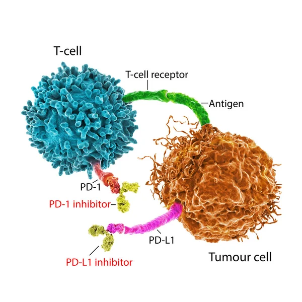
Immune Checkpoint Inhibitors In Cancer Treatment, 3D Illustration. Inhibitors Of PD-1 Receptor And PD-L1 Prevent The Tumour Cell From Binding To PD-1 And Enable The T Cell To Remain Active

Image, 6.99MB, 5352 × 5351 jpg
stock image The chemokine receptor 5 (CCR5) with a bound antagonist

The Chemokine Receptor 5 (CCR5) With A Bound Antagonist

Image, 4.59MB, 8000 × 6000 jpg
stock vector Mucosal immune system diagram. Mucous or gut associated lymphoid tissue. Medical vector illustration

Mucosal Immune System Diagram. Mucous Or Gut Associated Lymphoid Tissue. Medical Vector Illustration

Vector, 3.14MB, 5000 × 5000 eps
stock vector The life cycle of HIV. Stage 5 - Ribosomes lymphocyte cells synthesize proteins in the virus RNA. World AIDS Day.

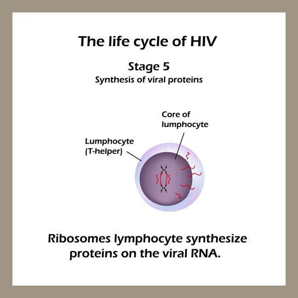
The Life Cycle Of HIV. Stage 5 - Ribosomes Lymphocyte Cells Synthesize Proteins In The Virus RNA. World AIDS Day.

Vector, 4.11MB, 5000 × 5000 eps
Page 1 >> Next