
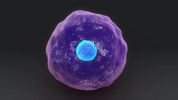
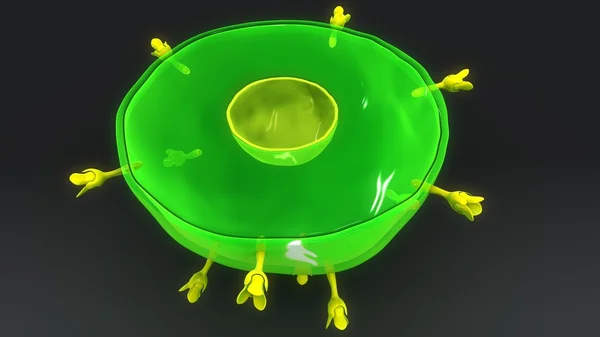
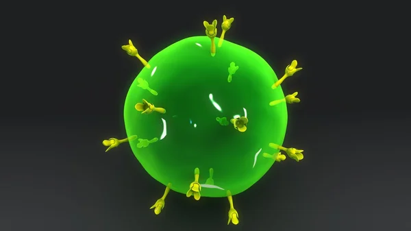
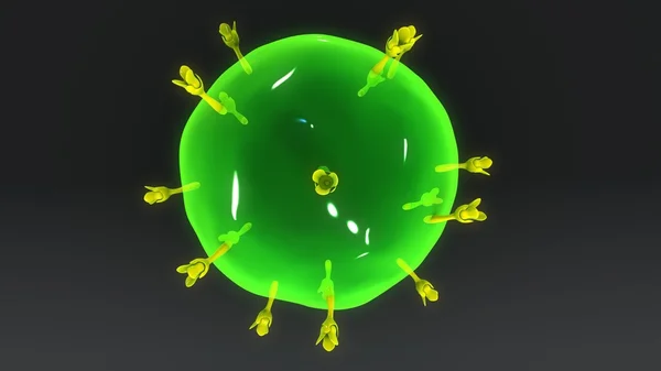
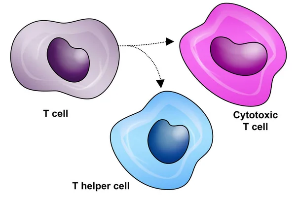
Stock image T Helper Cell
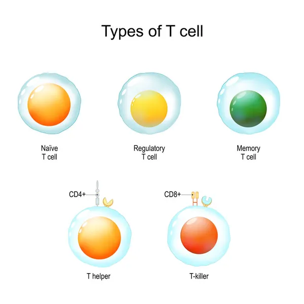
Types Of T Cell. From Naive And Memory Cells To T Helper And T-killer. Immunology Infographic. Vector Illustration
Vector, 13.06MB, 4444 × 4445 eps
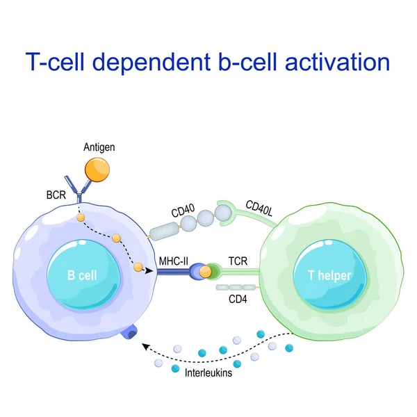
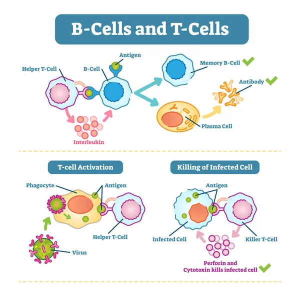
T-cell Dependent B-cell Activation. B Lymphocyte Binds An Antigen, Receive Help From A T Helper, And Differentiate Into A Plasma Cell That Secretes Of Antibodies. Receptors On Surface Of White Blood Cells. Human Immune System. Vector Poster
Vector, 1.05MB, 4444 × 4444 eps

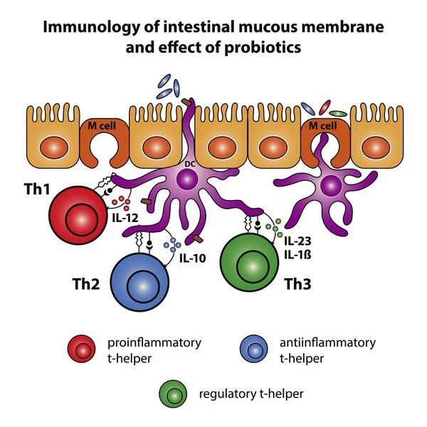
Mucosal Immune System Diagram. Mucous Or Gut Associated Lymphoid Tissue. Medical Vector Illustration
Vector, 3.14MB, 5000 × 5000 eps
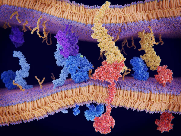
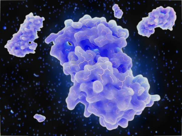
PD-1 (red) Extends From The Surface Of A T-cell And Interacts With The Ligand Protein PD-L1 (yellow) From A Antigen Presenting Cell. Although The T-cell Has Been Activated Through The Interaction Of A T-cell Receptor (blue) And A MHC Protein (viole
Image, 18.32MB, 8000 × 6000 jpg
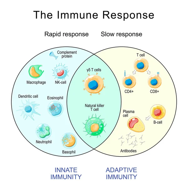
Immune Response. Rapid And Slow Response Of Adaptive And Innate Immunity And Antibody Activation. Cells Of The Immune System. Immunology Infographic. Vector Illustration
Vector, 2.28MB, 4444 × 4444 eps
Activation Of Leukocytes. T-cell Encounters Its Cognate Antigen On The Surface Of An Infected Cell. T-cells Direct And Regulate Immune Responses And Attack Infected Or Cancerous Cells. Cell-mediated Immunity. The Adaptive And Innate Immune System. Ve
Vector, 7.72MB, 7736 × 3000 eps

Lymphocytes. B Cell For Humoral Immunity. T-cell For Adaptive Immune Response. Vector Illustration. Poster
Vector, 12.91MB, 4444 × 4444 eps

Innate Immunity From Fever And Complement System (protein For Holes In The Plasma Membrane), To Macrophage, NK And Dendritic Cells. Adaptive Immunity From Antibodies And Plasma Cell To B-cell, T Helper, T-killer. Comparison And Difference
Vector, 2.86MB, 4444 × 4444 eps
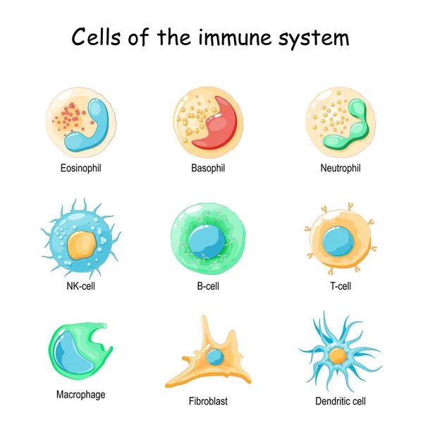
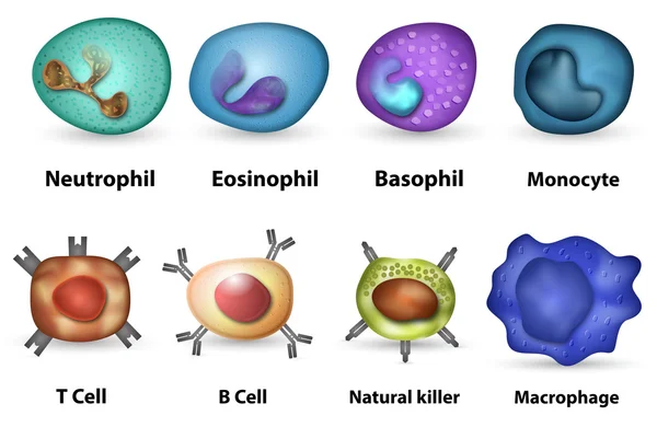
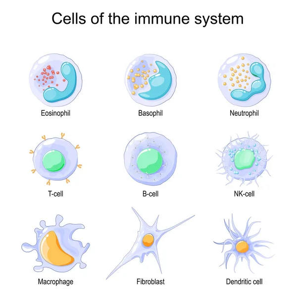
Cells Of The Immune System. White Blood Cells Or Leukocytes: Eosinophil, Neutrophil, Basophil, Macrophage, Fibroblast, And Dendritic Cell. Vector Diagram
Vector, 1.83MB, 4444 × 4444 eps
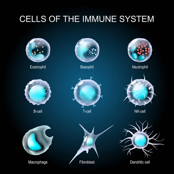
Cells Of The Immune System. White Blood Cells Or Leukocytes Eosinophil, Neutrophil, Basophil, Macrophage, Fibroblast, And Dendritic Cell. Set Of Transparent Realistic Cells On A Dark Background. Vector Illustration
Vector, 5.41MB, 4444 × 4444 eps

Types Of Lymphocytes. T And B Cell. Human Adaptive Immune System. Immune Response And Component Of Humoral Immunity. Antibody, Plasma Cell, T Helper, And T-killer. Vector Illustration
Vector, 1.63MB, 4444 × 4444 eps
Cells Of The Immune System. White Blood Cells Or Leukocytes Eosinophil, Neutrophil, Basophil, Macrophage, Fibroblast, And Dendritic Cell. Vector Illustration
Vector, 1.73MB, 4444 × 4445 eps

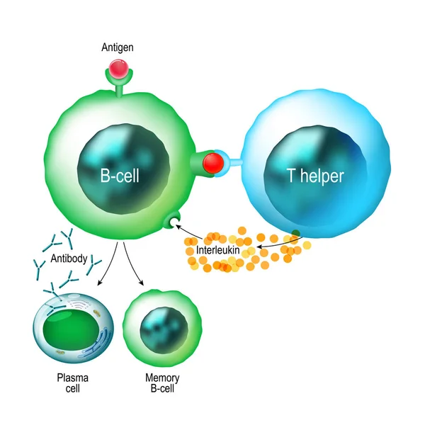
Immune Response And Antigen Presentation. Humoral Immunity And Antibody Production. B-cell Activation. Vector Poster For Education
Vector, 10.45MB, 4444 × 4444 eps

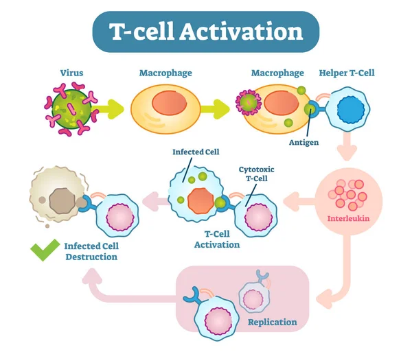
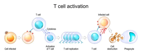
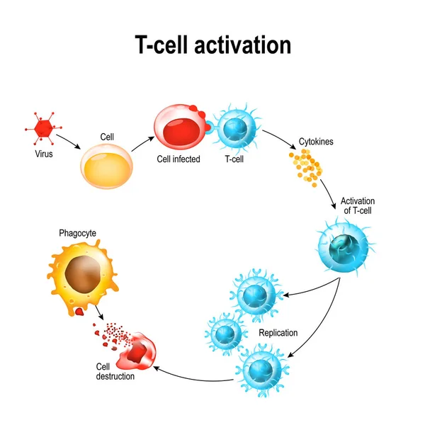
Immune Response And Antigen Presentation. Cell-mediated Immunity Is An Immune Response That Does Not Involve Antibodies. T-cell Activation. Vector Poster
Vector, 10.2MB, 4444 × 4444 eps
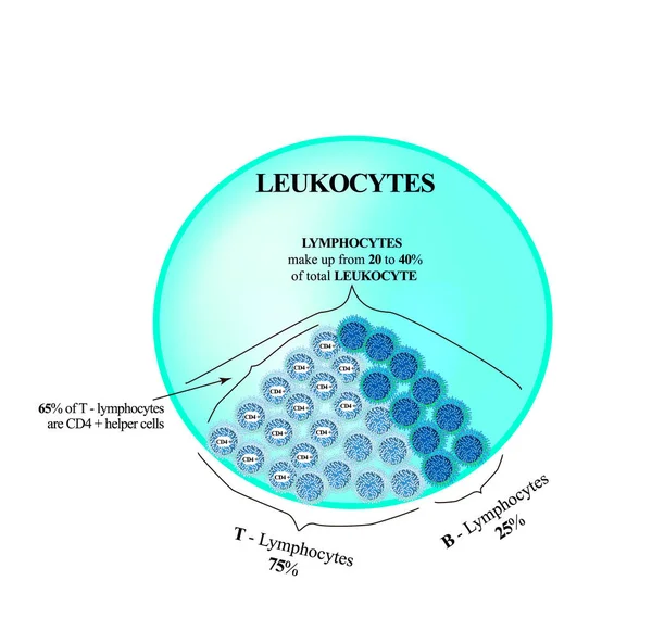
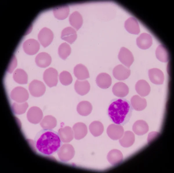
Lymphocytes Make Up From 20 To 40 Percent Of The Total Number Of Leukocytes. T Lymphocytes And B Lymphocytes. Cell Killers. Immunity Helper Cells. Infographics. Vector Illustration
Vector, 9.44MB, 5163 × 5000 eps

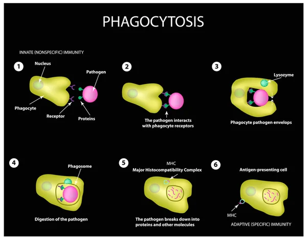
Innate Immunity. Adaptive Specific . Phagocytosis. Infographics. Vector
Vector, 3.99MB, 5000 × 3911 eps
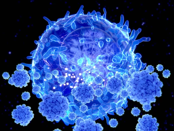
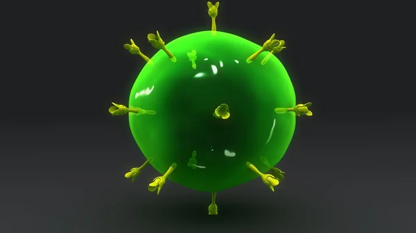
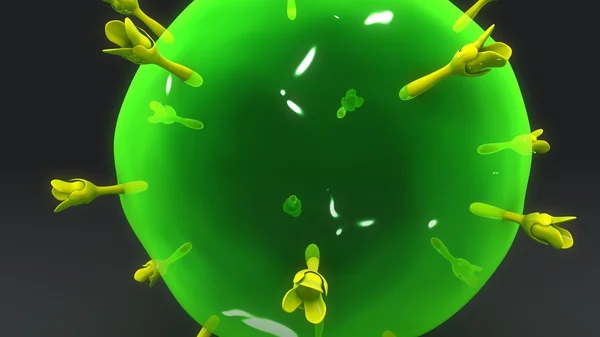
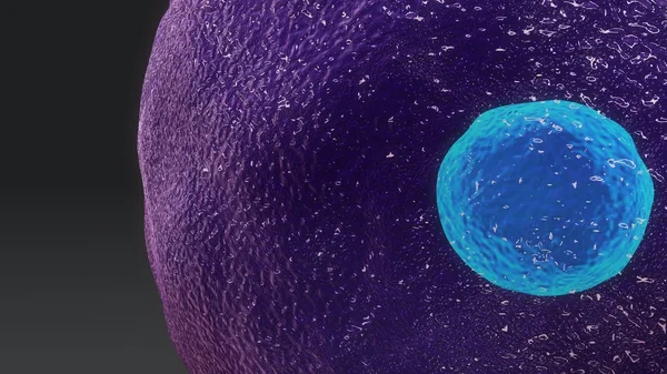
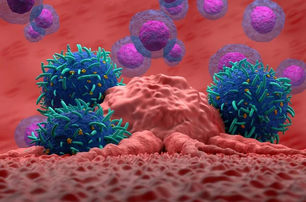
T Cell Targeting SARS-CoV-2 Viruses. T Cells Play A Key Role In The Development Of Long-term Immunity. To Coronavirus
Image, 8.67MB, 8000 × 6000 jpg
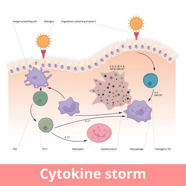
Cytokine Storm. Hypercytokinemia During Which The Immune System Causes A Release Of Cytokines With Help Of Macrophages, T Helper Cells, Neutrophils.
Vector, 6.7MB, 6250 × 6250 eps
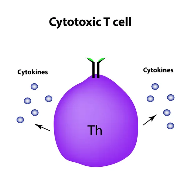
Cytotoxic Cells. Cytokines. Cell Immunity. Infographics. Vector Illustration
Vector, 1.39MB, 5000 × 5000 eps
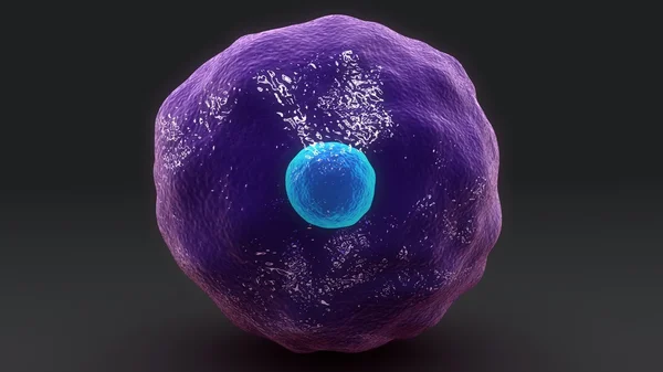
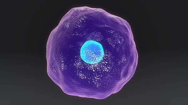
3d Computer Illustration Of A Dendritic Cell. They Areantigen-presenting Cells Of The Immune System. Their Main Function Is To Process Antigen Material And Present It On The Cell Surface To The T Cells Of The Immune System. They Are Messengers Betwe
Image, 5.2MB, 8000 × 6000 jpg

Innate Immunity. Adaptive Specific . Phagocytosis. Infographics. Vector Illustration
Vector, 1.48MB, 5000 × 4734 eps
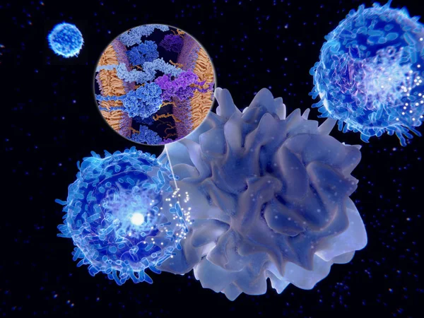
Dendritic Cells Present Antigens (green) To Lymphocytes Through Their Membran Bound MHC-molecules (violet). CD4 Molecules (light Blue) Bind To Other Portions Of The MHC, Strengthening The Interaction.
Image, 10.24MB, 8000 × 6000 jpg
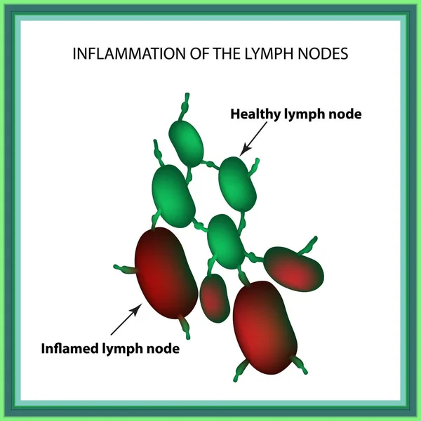
Inflammation Of The Lymph Nodes. Infographics. Vector Illustration On Isolated Background
Vector, 1.13MB, 5000 × 5000 eps

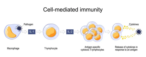
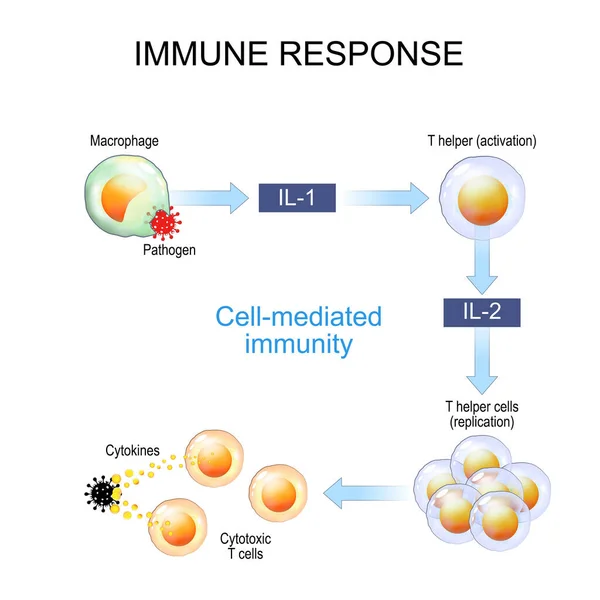
Immune Response. Cell-mediated Immunity. Activation Of Phagocytes, Antigen-specific Cytotoxic T-lymphocytes, And The Release Of Cytokines In Response To An Antigen. Vector Poster For Educatio
Vector, 11.15MB, 7000 × 2734 eps
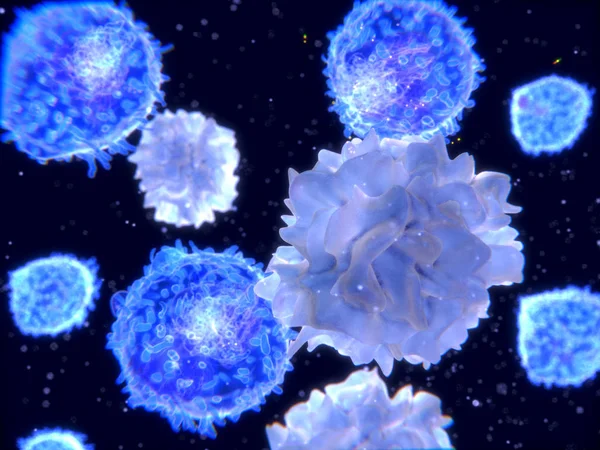
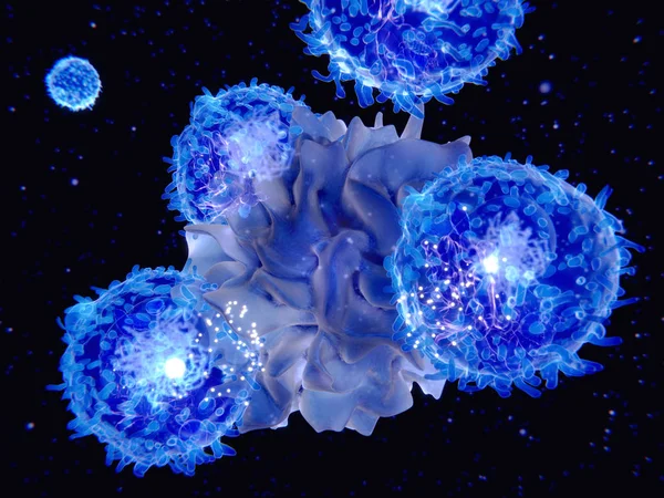
T-lymphocytes And Dendritic Cells, 3D-rendering; Dendritic Cells Are Antigen-presenting Cells Of The Immune System. They Process Antigen Material And Present It On The Cell Surface To The T-cells. Illustration
Image, 2.23MB, 4000 × 3000 jpg

T-cell Receptor In Complex With The MHC Class II-peptide Complex. The Antigen (light Green) Is A Peptide From A Tumor Cell, Bacteria Or Virus. Different Stages Of The Interaction. 3D-Rendering. Illustration
Image, 7.31MB, 8000 × 6000 jpg
Page 1 >> Next