
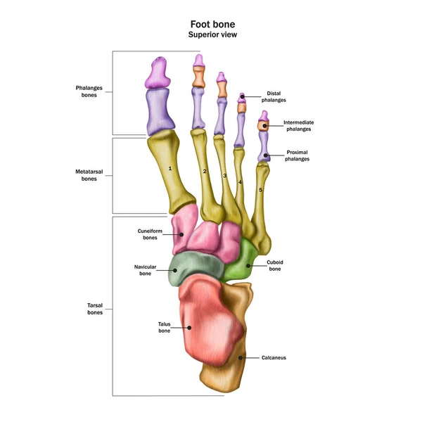
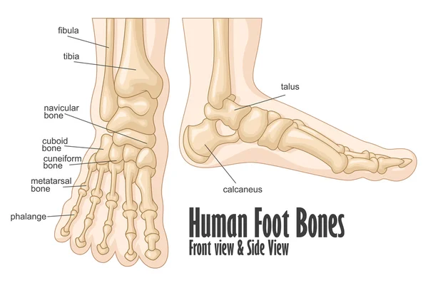
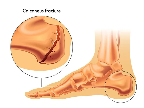
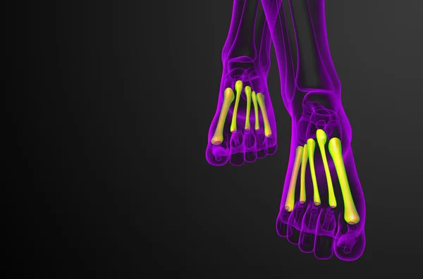
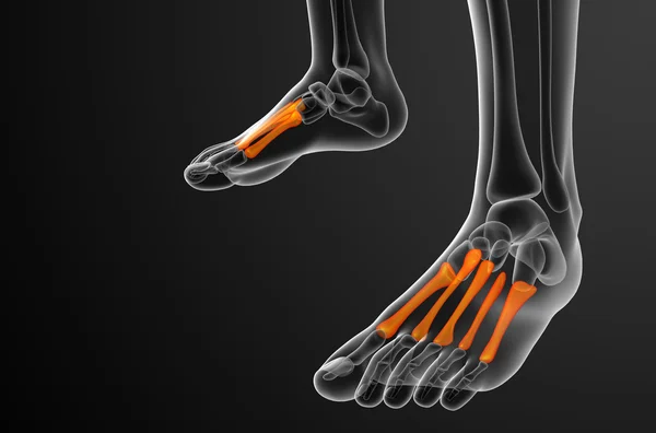
Stock image Tarsal

Ankle Pain From Instability, Arthritis, Gout, Tendonitis, Fracture, Nerve Compression (tarsal Tunnel Syndrome), Infection And Poor Structural Alignment Of Leg Or Foot In Ageing Patient With Doctor
Image, 4.27MB, 3472 × 2314 jpg

Doctor Holding A Digital Tablet With X-ray And Pain On The Ankle Of The Foot
Image, 7.44MB, 6000 × 4000 jpg

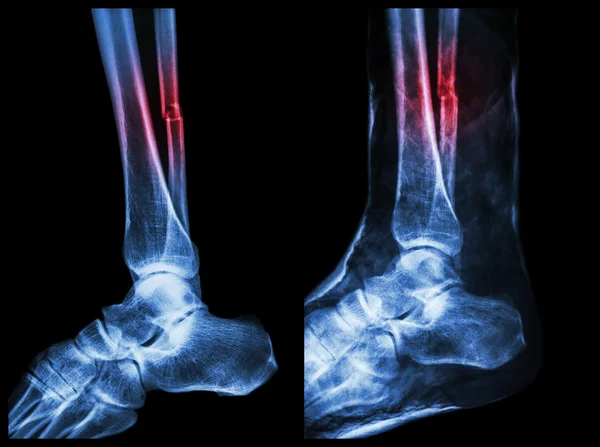
Left Image : Fracture Shaft Of Fibula (calf Bone) , Right Image : It Was Splinted With Plaster Cast
Image, 11.19MB, 7142 × 5331 jpg
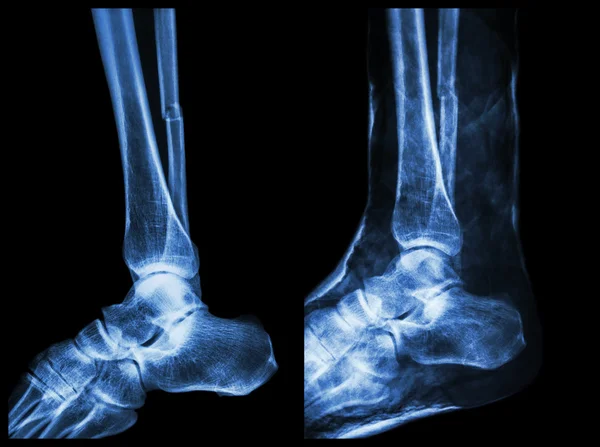
Left Image : Fracture Shaft Of Fibula (calf Bone) , Right Image : It Was Splinted With Plaster Cast
Image, 13.26MB, 7142 × 5331 jpg

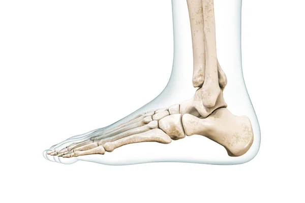
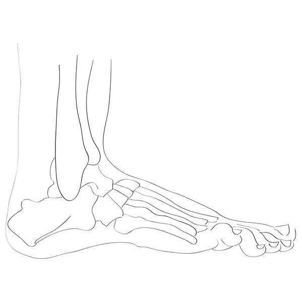
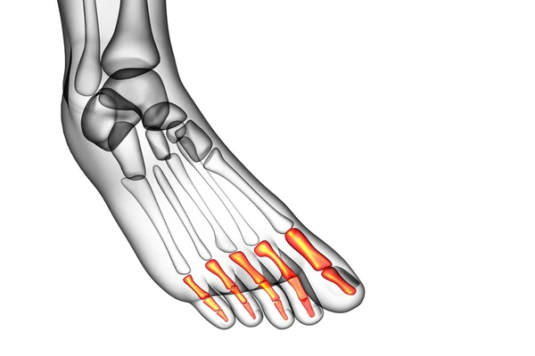
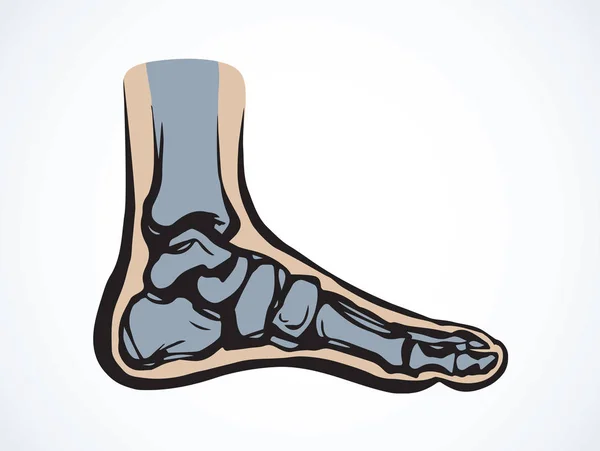
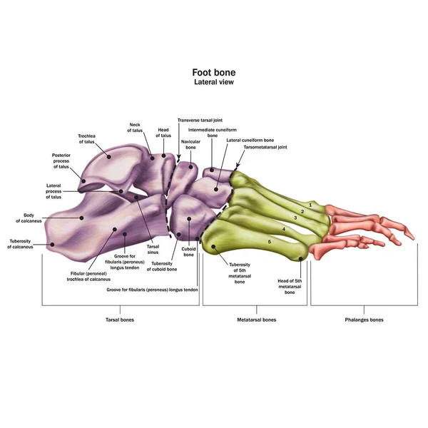
Foot Bones Lateral View With Body Contours 3D Rendering Illustration Isolated On White With Copy Space. Human Skeleton And Ankle Anatomy, Medical Diagram, Osteology, Skeletal System Concepts.
Image, 1.64MB, 4000 × 2667 jpg
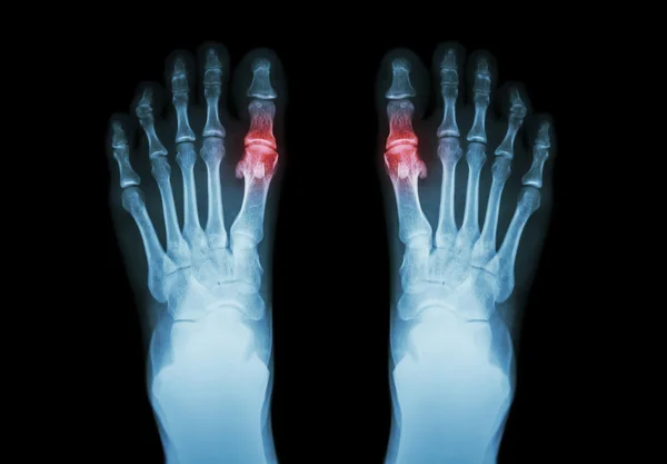
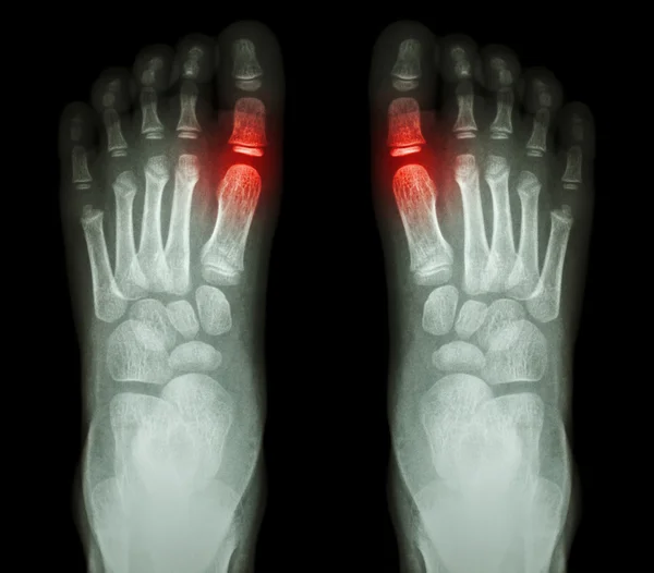
Gout , Rheumatoid Arthritis ( Film X-ray Both Foot And Arthritis At First Metatarsophalangeal Joint ) ( Medicine And Science Background )
Image, 2.86MB, 4684 × 3257 jpg


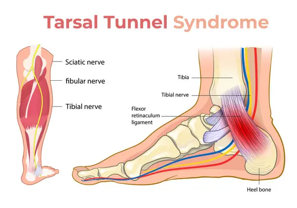
Neurological Diagnosis Of Tunnel Syndrome. Neurologist Directory, Where Is Printed Diagnosis Tunnel Syndrome, Lies On Workplace With MRI Image And Neurological Diagnostic Tools Close Up
Image, 5.96MB, 6016 × 4000 jpg

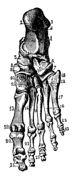
The Tarsal Bones Of The Foot Are Located In The Midfoot And The Hind Foot Areas Of The Human Foot, Vintage Line Drawing Or Engraving Illustration.
Vector, 3.86MB, 3978 × 9719 eps
Left Image : Fracture Shaft Of Fibula (calf Bone) , Right Image : It Was Splinted With Plaster Cast
Image, 13.06MB, 7142 × 5331 jpg


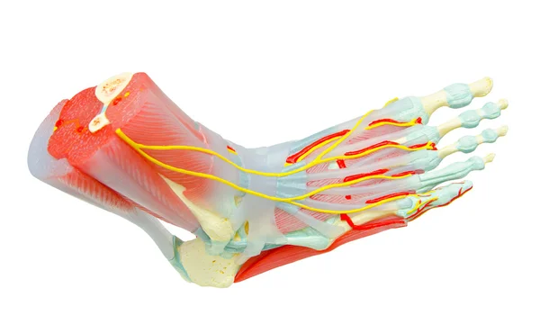
Human Anatomy. Nerves Of The Sole Of The Right Foot On A White Background. 3D Illustration
Image, 2.74MB, 4167 × 6471 jpg

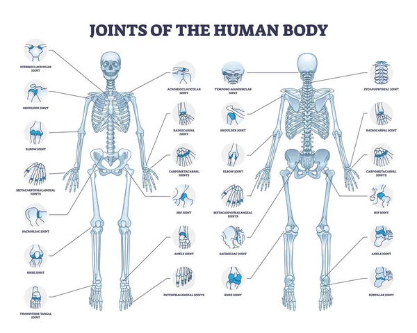
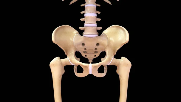
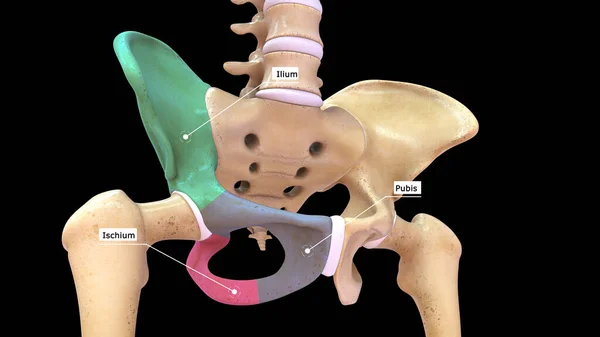
Joints Of Human Body With All Medical Parts Collection In Outline Diagram. Labeled Educational Scheme With Skeleton And Bone Connection Points Location Vector Illustration. Anatomical Explanation.
Vector, 19.7MB, 4800 × 3840 eps


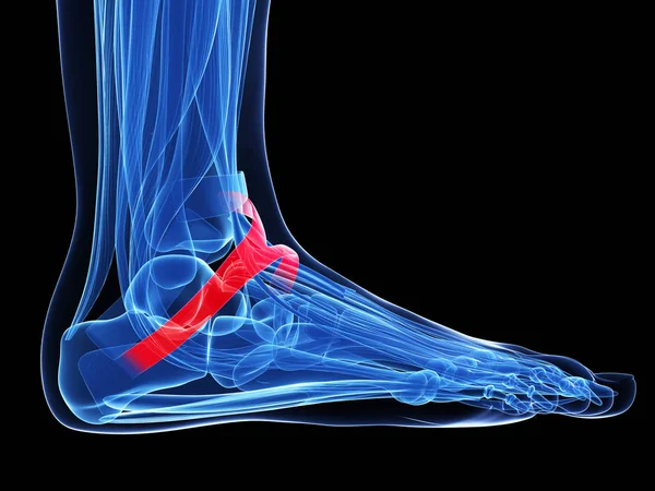
Foot And Ankle Pain, Conceptual 3D Illustration. Human Anatomy. A Male Body In Pose Showing Foot Pain With Highlighted Skeleton And Isolated Skeleton
Image, 9.39MB, 11360 × 5000 jpg

Doctor Holding A Digital Tablet With X-ray With Pain On The Toe Of The Foot
Image, 7.37MB, 6000 × 4000 jpg


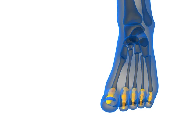
"Rheumatoid Arthritis , Gouty Arthritis" X-ray Child's Foots And Arthritis At Metatarsophalangeal Joint (Big Toe Area)
Image, 9.5MB, 5700 × 5000 jpg

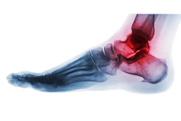
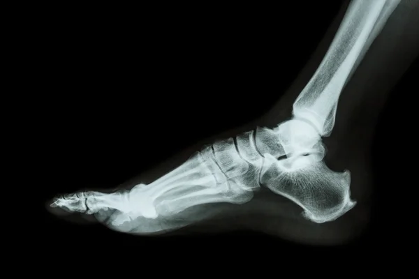
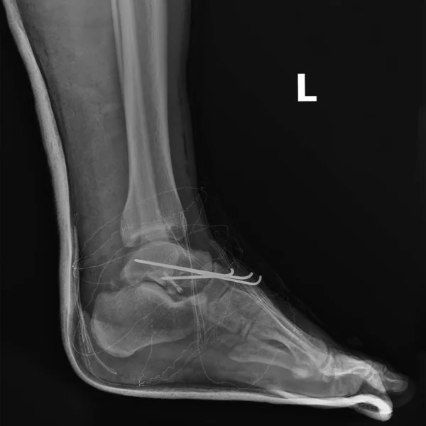
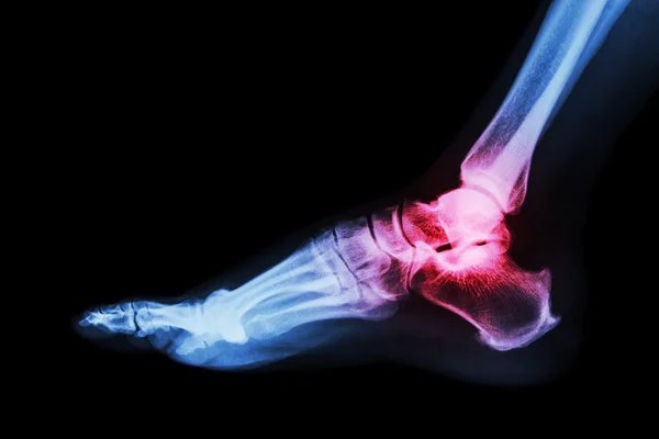
Arthritis Of Ankle . X-ray Of Foot . Lateral View . Invert Color Style .
Image, 4.87MB, 5184 × 3456 jpg
Page 1 >> Next