
Stock image Tectorial
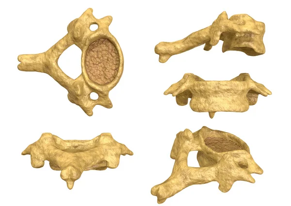
Morphology Of The Cervical Vertebra, Sixth Cervical Vertebra, Multiple Angles And Views
Image, 3.41MB, 3508 × 2480 jpg

The Ligaments Of The Median Atlantoaxial Joint. Atlas And Axis Ligaments. Cervical Spine, Vertebral Morphology, First And Second Cervical Vertebra, Cervical Vertebrae, Atlas, Axis, Atlantoaxial Joint, Superior View
Image, 4.03MB, 5906 × 4229 jpg

Light Micrograph Of A Cross Section Of The Cochlea Of The Inner Ear Showing From Top To Bottom: Vestibular, Cochlear And Tympanic Ducts Or Scala , Separated By Reissner Membrane And Basilar Membrane. The Cochlear Duct Shows, From Right To Left, The S
Image, 4.37MB, 3840 × 3072 jpg
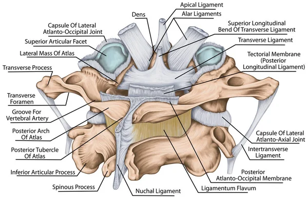
The Ligaments Of The Median Atlantoaxial Joint. Atlas And Axis Ligaments. Cervical Spine, Vertebral Morphology, First And Second Cervical Vertebra, Cervical Vertebrae, Atlas, Axis, Atlantoaxial Joint, Posterosuperior View
Image, 5.91MB, 5906 × 3830 jpg
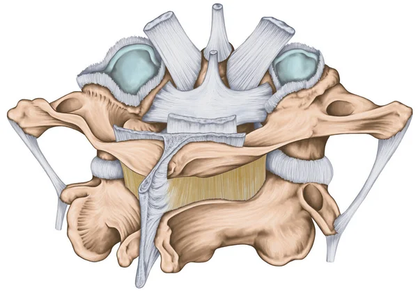
The Ligaments Of The Median Atlantoaxial Joint. Atlas And Axis Ligaments. Cervical Spine, Vertebral Morphology, First And Second Cervical Vertebra, Cervical Vertebrae, Atlas, Axis, Atlantoaxial Joint, Posterosuperior View
Image, 5.87MB, 5906 × 4183 jpg
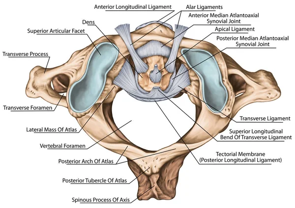
The Ligaments Of The Median Atlantoaxial Joint. Atlas And Axis Ligaments. Cervical Spine, Vertebral Morphology, First And Second Cervical Vertebra, Cervical Vertebrae, Atlas, Axis, Atlantoaxial Joint, Superior View
Image, 4.75MB, 5906 × 4181 jpg

Healthy And Damaged Hair Cells Inside Cochlea. Noise-induced Hearing Loss. Tinnitus.
Vector, 4.64MB, 6000 × 4500 eps
Ligaments And Joints Of The Cervical Vertebrae And The Occipital Bone. Back View. Vector Illustration
Vector, 2.91MB, 6010 × 4167 eps

Healthy And Damaged Hair Cells Inside Cochlea. Noise-induced Hearing Loss. Tinnitus.
Vector, 7.2MB, 6000 × 4500 eps

Light Micrograph Of A Cross Section Of The Cochlea Scala Media Or Cochlear Duct Showing From Right To Left: Limbus Spiralis With Tectorial Membrane And Reissner Membrane, Organ Of Corti With Tunnel Of Corti And Hair Cells, And The Stria Vascularis.
Image, 5.45MB, 3840 × 3072 jpg
Page 1 >> Next