photo free
Home Images Vectors Videos

Stock image Trichinellosis

stock vector Trichomonas vaginalis structure. Trichomoniasis. Urogenital infection. Infographics. Vector illustration on isolated background.

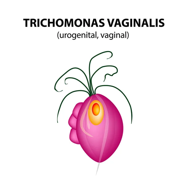
Trichomonas Vaginalis Structure. Trichomoniasis. Urogenital Infection. Infographics. Vector Illustration On Isolated Background.

Vector, 1.31MB, 5000 × 5000 eps
stock image Trichinella spiralis larvae in muscle tissue under the microscope.

Trichinella Spiralis Larvae In Muscle Tissue Under The Microscope.

Image, 1.79MB, 4500 × 3000 jpg
stock vector Trichomonas vaginalis, Trichomonas vaginalis diagram, Trichomonas vaginalis anatomy, Trichomonas vaginalis structure

Trichomonas Vaginalis, Trichomonas Vaginalis Diagram, Trichomonas Vaginalis Anatomy, Trichomonas Vaginalis Structure

Vector, 26.72MB, 5622 × 3922 eps
stock image Stop trichinosis. Syringe is filled with injection. Syringe and vaccine

Stop Trichinosis. Syringe Is Filled With Injection. Syringe And Vaccine

Image, 3.25MB, 4400 × 2942 jpg
stock image The diagnosis of trichinosis

The Diagnosis Of Trichinosis

Image, 5.05MB, 4920 × 3384 jpg
stock image Trichinella spiralis - parasitic worm microscope

Trichinella Spiralis - Parasitic Worm Microscope

Image, 7.85MB, 3333 × 2500 jpg
stock image Trichinella spiralis larvae in muscle tissue under the microscope.

Trichinella Spiralis Larvae In Muscle Tissue Under The Microscope.

Image, 1.51MB, 4500 × 3000 jpg
stock image View in the microscope on Trichomonas.

View In The Microscope On Trichomonas.

Image, 1.84MB, 2327 × 2300 jpg
stock image raw pork belly with rind

Raw Pork Belly With Rind

Image, 9.78MB, 7360 × 4912 jpg
stock image 3D illustration and micrograph, transverse section, of cyst in muscle containing helminth Trichinella spiralis, nematode larval cyst in muscle tissue, transmitted by ingestion of undercooked meat

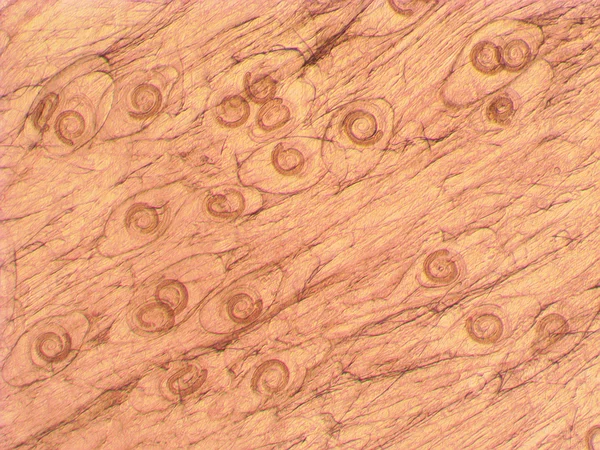
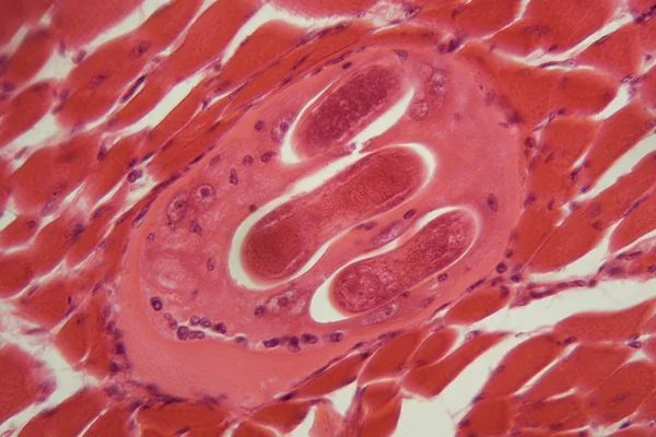
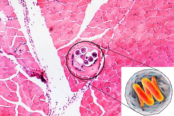
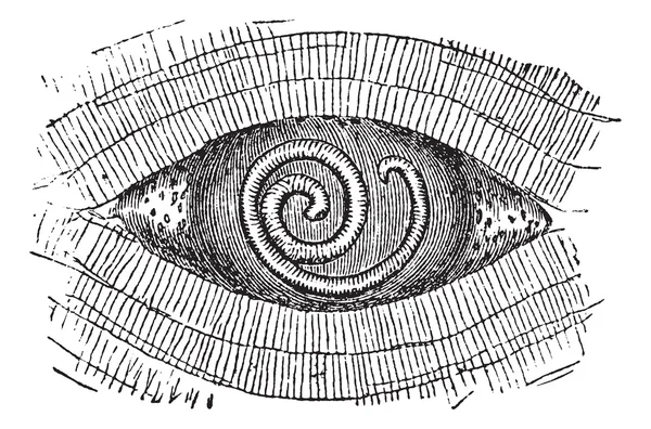
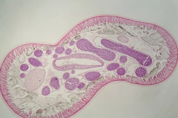
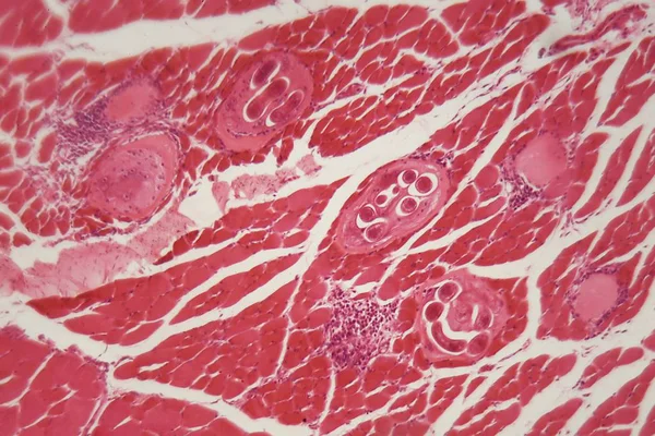
3D Illustration And Micrograph, Transverse Section, Of Cyst In Muscle Containing Helminth Trichinella Spiralis, Nematode Larval Cyst In Muscle Tissue, Transmitted By Ingestion Of Undercooked Meat

Image, 3.9MB, 2854 × 1903 jpg
stock image Positive result of blood test for trichinosis. Test tube with a blood test in the doctor's hands. Medical concept.

Positive Result Of Blood Test For Trichinosis. Test Tube With A Blood Test In The Doctor's Hands. Medical Concept.

Image, 5.03MB, 3876 × 2672 jpg
stock image Trichinosis - diagnosis written on a white piece of paper. Syringe and vaccine with drugs

Trichinosis - Diagnosis Written On A White Piece Of Paper. Syringe And Vaccine With Drugs

Image, 10.97MB, 5184 × 3456 jpg
stock vector Trichinosis, vintage engraving

Trichinosis, Vintage Engraving

Vector, 0.99MB, 5634 × 9508 eps
stock vector Fig. 2. Trichinella adult female, magnified 150 times, vintage e

Fig. 2. Trichinella Adult Female, Magnified 150 Times, Vintage E

Vector, 3.53MB, 6633 × 9866 eps
stock image Trichinella spiralis larvae in muscle tissue under the microscope.

Trichinella Spiralis Larvae In Muscle Tissue Under The Microscope.

Image, 1.86MB, 4500 × 3000 jpg
stock image A veterinarian is examining sample of meat, pork lung tissue, on trichinosis, looking on glass tiles under an electric microscope at outdoor laboratory.

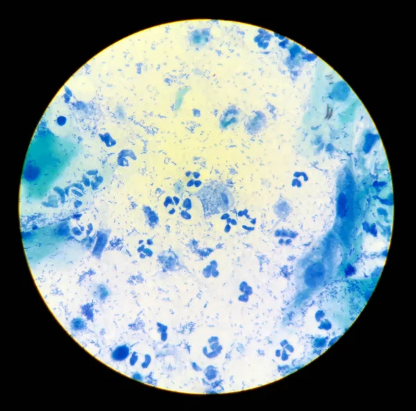
A Veterinarian Is Examining Sample Of Meat, Pork Lung Tissue, On Trichinosis, Looking On Glass Tiles Under An Electric Microscope At Outdoor Laboratory.

Image, 7.32MB, 5184 × 3456 jpg
stock image Trichinosis - diagnosis written on a white piece of paper. Syringe and vaccine with drugs

Trichinosis - Diagnosis Written On A White Piece Of Paper. Syringe And Vaccine With Drugs

Image, 10.5MB, 5184 × 3456 jpg
stock vector Oral Trichomonas structure. Trichomoniasis. Urogenital infection. Infographics. Vector illustration on isolated background.

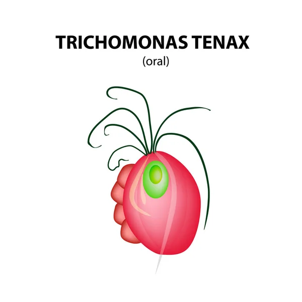
Oral Trichomonas Structure. Trichomoniasis. Urogenital Infection. Infographics. Vector Illustration On Isolated Background.

Vector, 1.58MB, 5000 × 5000 eps
stock vector Sexually transmitted infection. Close-up of causative agents of Venereal disease: Ureaplasma, Trichomonas, Candida, Treponema, Chlamydiales, Neisseria gonorrhoeae, Herpes virus, HIV, papillomavirus

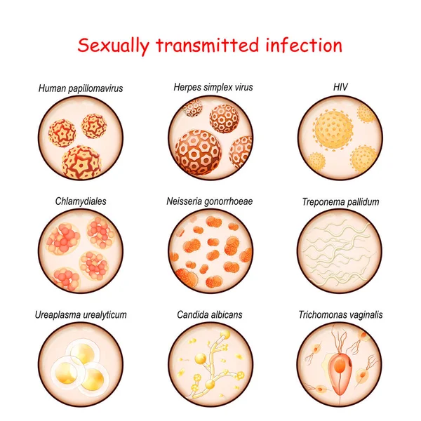
Sexually Transmitted Infection. Close-up Of Causative Agents Of Venereal Disease: Ureaplasma, Trichomonas, Candida, Treponema, Chlamydiales, Neisseria Gonorrhoeae, Herpes Virus, HIV, Papillomavirus

Vector, 10.47MB, 4444 × 4444 eps
stock vector Trichomonas hominis- parasitic microscope

Trichomonas Hominis- Parasitic Microscope

Vector, 1.49MB, 4000 × 4000 eps
stock vector Pork Roundworm or Trichinella spiralis, vintage engraving

Pork Roundworm Or Trichinella Spiralis, Vintage Engraving

Vector, 1.81MB, 6633 × 4384 eps
stock image pork belly on the white background

Pork Belly On The White Background

Image, 6.3MB, 7360 × 4912 jpg
stock vector Trichomonas vaginal. Infographics. Vector illustration on isolated background.

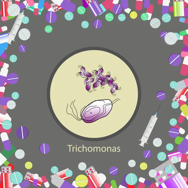
Trichomonas Vaginal. Infographics. Vector Illustration On Isolated Background.

Vector, 8.42MB, 5208 × 5208 eps
stock image Parasitic nematode worm (Ascaris sp.) under the microscope

Parasitic Nematode Worm (Ascaris Sp.) Under The Microscope

Image, 2.83MB, 4500 × 3000 jpg
stock vector Infection caused by eating of undercooked pork meat infected with parasitic larvae

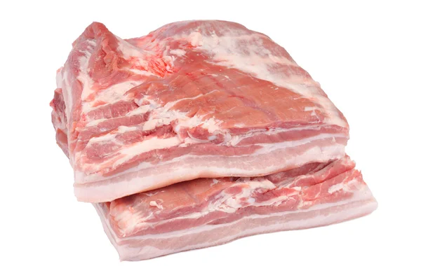
Infection Caused By Eating Of Undercooked Pork Meat Infected With Parasitic Larvae

Vector, 0.53MB, 4168 × 4168 eps
stock vector Types Trichomonads. Intestinal, oral, vaginal trichomonas structure. Trichomoniasis. Urogenital infection. Infographics. Vector illustration on isolated background.

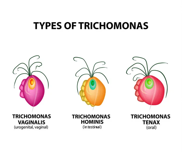
Types Trichomonads. Intestinal, Oral, Vaginal Trichomonas Structure. Trichomoniasis. Urogenital Infection. Infographics. Vector Illustration On Isolated Background.

Vector, 2.16MB, 5000 × 4174 eps
stock image Trichinella spiralis larvae in muscle tissue under the microscope.

Trichinella Spiralis Larvae In Muscle Tissue Under The Microscope.

Image, 1.87MB, 4500 × 3000 jpg
stock vector Fig. 1. Piece of flesh pork trichina - grown, vintage engraving.

Fig. 1. Piece Of Flesh Pork Trichina - Grown, Vintage Engraving.

Vector, 11.42MB, 6633 × 6527 eps
Page 1 >> Next