
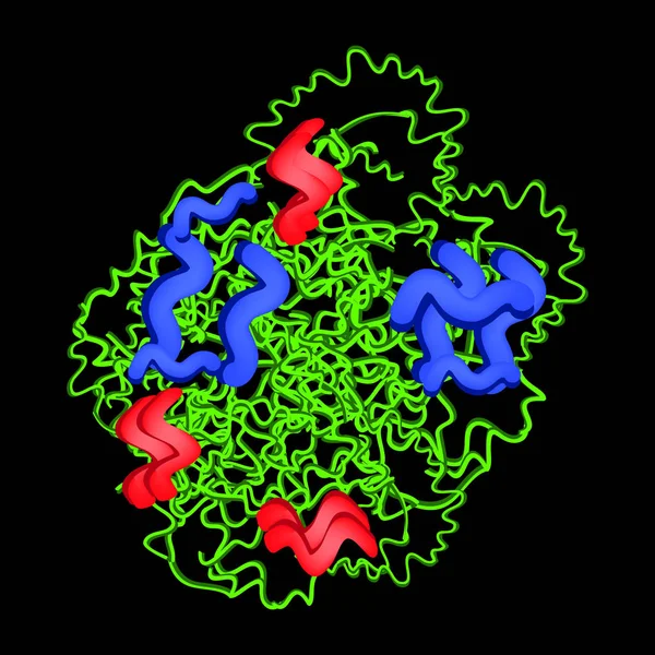
Stock image Trypsin

Trypsin Digestive Enzyme Molecule (human). 3D Illustration. Enzyme That Contributes To The Digestion Of Proteins In The Digestive System.
Image, 6.01MB, 8000 × 6000 jpg
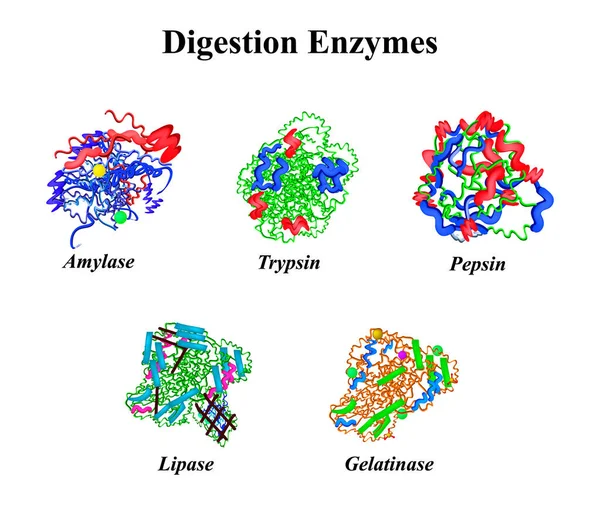
Digestion Enzymes Set. Chemical Molecular Formula. Amylase, Trypsin, Gelatinase, Pepsin, Lipase. Infographics. Vector Illustration On Isolated Background.
Vector, 7.37MB, 5000 × 4231 eps
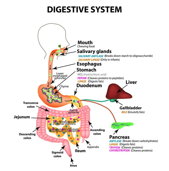
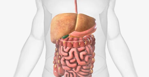
The Human Digestive System. Anatomical Structure. Digestion Of Carbohydrates, Fats And Proteins. Enzymes Of The Gastrointestinal Tract, Pancreas, Liver, Gallbladder. Metabolism. Infographics. Vector.
Vector, 3.01MB, 5000 × 5050 eps
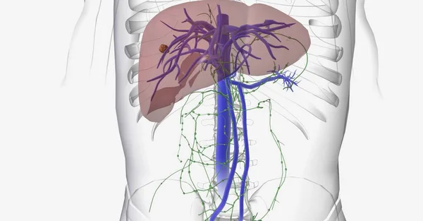
The Most Common Type Of Cancer That Begins In The Liver Is Called Hepatocellular Carcinoma (HCC). 3D Rendering
Image, 13.41MB, 11115 × 5802 jpg
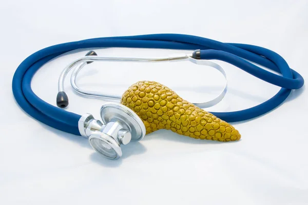
Pancreas Gland Is Next To Stethoscope And His Head, Symbolizing Process Of Diagnosis Of Pancreas Diseases, Identify Causes Of Increase Or Decrease Production Of Hormones And Other Endocrine Problems
Image, 5.95MB, 6000 × 4000 jpg
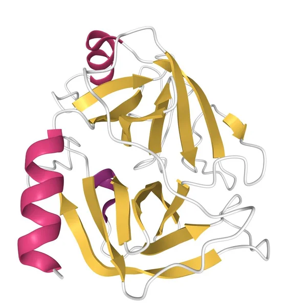
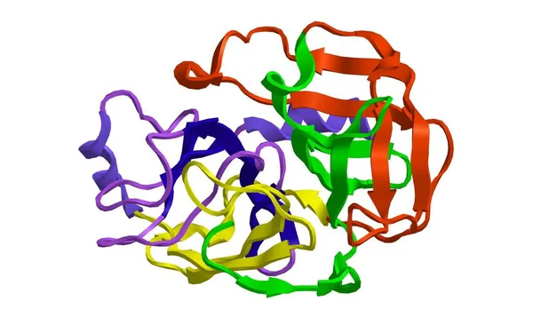
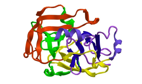
Structure Of Human Trypsin IV (brain Trypsin), 3D Cartoon Model Isolated, White Background
Image, 1.82MB, 4096 × 4096 jpg

Digestion Of Protein. Breaking The Complex Molecule First Into Peptides Then Into Individual Amino Acids. The Pepsins Are Enzymes Secreted By The Stomach That Breaks Down Proteins. Vector Illustration
Vector, 5.26MB, 6000 × 3321 eps

Trypsin Molecular Chemical Formula. Enzyme Of The Pancreas. Infographics. Vector Illustration On Isolated Background
Vector, 1.77MB, 5000 × 5000 eps
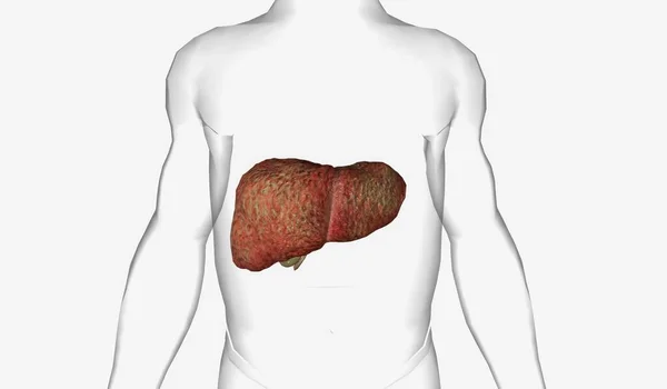
Nonalcoholic Fatty Liver Disease (NAFLD) Is A Condition In Which Excess Fat Cells Are Stored In The Liver. 3D Rendering
Image, 16.57MB, 11115 × 5802 jpg
Trypsin Molecular Chemical Formula. Enzyme Of The Pancreas. Infographics. Vector Illustration On Black Background
Vector, 1.76MB, 5000 × 5000 eps


Anatomical Models Of Pancreas And Stomach Lie Close One By One On White Background. Concept Of Digestive System And Organs, Where Processing And Preparation Of Food For Further Absorption In Intestine
Image, 4.7MB, 6016 × 4000 jpg
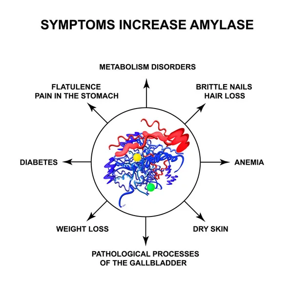
Symptoms Of Increased Amylase. The Enzyme Amylase. Infographics. Vector Illustration On Isolated Background.
Vector, 2.14MB, 5000 × 4830 eps
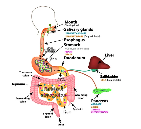
The Human Digestive System. Anatomical Structure. Digestion Of Carbohydrates, Fats And Proteins. Enzymes Of The Gastrointestinal Tract, Pancreas, Liver, Gallbladder. Metabolism. Infographics. Vector.
Vector, 2.89MB, 5000 × 4572 eps

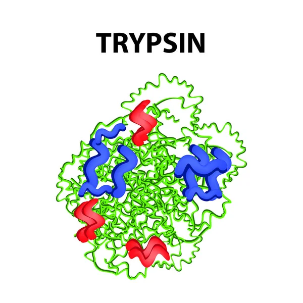
Trypsin Molecular Chemical Formula. Enzyme Of The Pancreas. Infographics. Vector Illustration On Isolated Background
Vector, 1.71MB, 5000 × 5000 eps

Arginine As A Complex Subject, Related To Important Topics. Pictured As A Puzzle And A Word Cloud Made Of Most Important Ideas And Phrases Related To Arginine.
Image, 2.91MB, 7680 × 4320 jpg
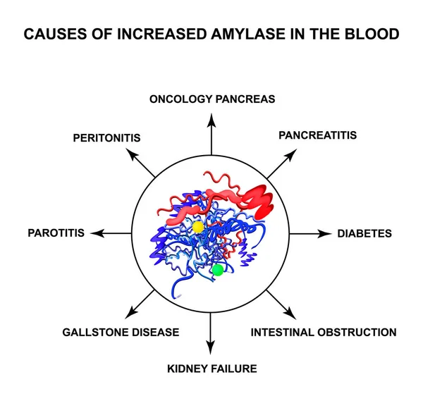
Causes Of Increased Amylase In The Blood. The Enzyme Amylase. Infographics. Vector Illustration On Isolated Background
Vector, 2.12MB, 5000 × 4830 eps
PBC Is Characterized By Autoimmune Destruction Of The Small And Medium Sized Bile Ducts In The Liver. 3D Rendering
Image, 3.41MB, 7191 × 4200 jpg
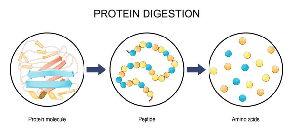
Protein Digestion. Enzymes Proteases And Peptidases Are Digestion Breaks The Protein Into Smaller Peptide Chains And Into Single Amino Acids, Which Are Absorbed Into The Blood. Vector Illustration
Vector, 1.22MB, 6742 × 3000 eps
Page 1 >> Next