
Stock image Tuberosity
Pelvis Bones Of Pelvic Girdle, Vector Sketch Of Human Anatomy And Medicine. Bones And Joints Structure Of Skeleton Hips, Sacrum, Femur And Coccyx, Sacral Promontory, Pubic Arch And Iliac Spine
Vector, 2.56MB, 5957 × 5036 eps
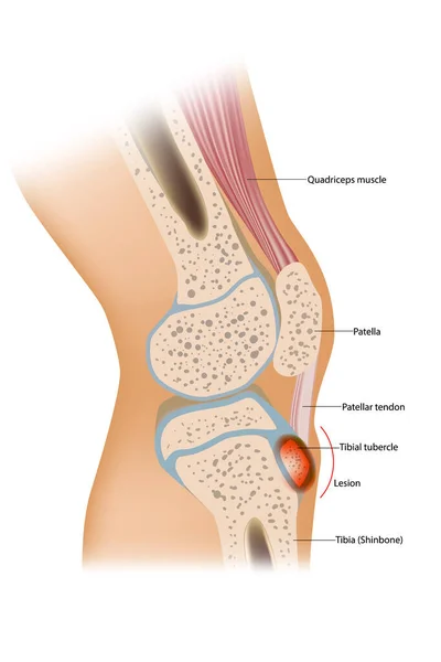
Osgood Schlatter Disease Or OSD Is Inflammation Of The Patellar Ligament At The Tibial Tuberosity
Vector, 4.44MB, 3900 × 5850 eps
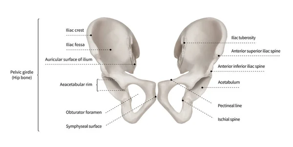
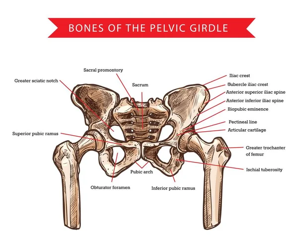
Infographic Diagram Of Human Hip Bone Or Pelvic Girdle Anatomy System Anterior View- 3D- Human Anatomy- Medical Diagram- Educational And Human Body Concept- Isolated On White Background
Image, 8.54MB, 15000 × 7536 jpg

Gluteus Medius Muscle With Human Hip And Groin Anatomy Outline Diagram
Vector, 9.26MB, 4500 × 4018 eps

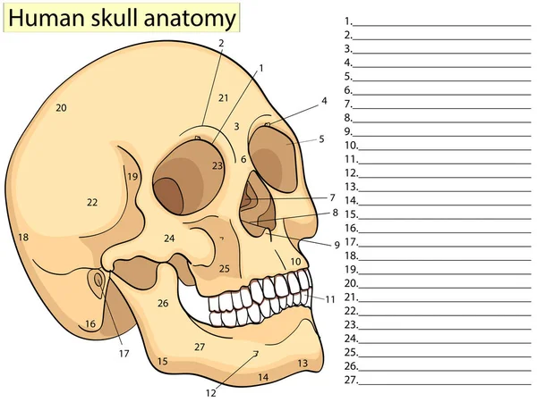
Medical Education Chart Of Biology Human Skull Diagram. Vector. Front Aspect White Background Basic Medical Education
Vector, 1.21MB, 5333 × 4000 eps

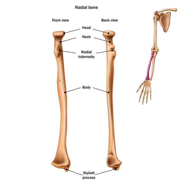
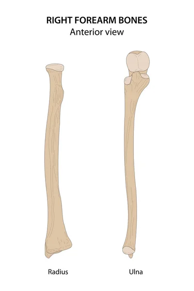
Radius And Ulna Bone Anatomy With Arm Skeletal Structure Outline Diagram. Labeled Educational Scheme With Upper Body Parts And Hand Long Bones Vector Illustration. Detailed Physiological Description.
Vector, 9.7MB, 5000 × 3750 eps

Stress Fracture. Marching Fracture Of The Foot. Fracture Of The Metatarsal Bones In The Foot. Anatomical Structure Of The Foot. Skeleton. Broken Bones. Vector Illustration On Isolated Background.
Vector, 2.69MB, 5000 × 5000 eps
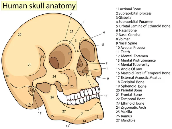
Medical Education Chart Of Biology Human Skull Diagram. Vector. Front Aspect White Background Basic Medical Education
Vector, 1.1MB, 5333 × 4000 eps
Medical Education Chart Of Biology Human Skull Diagram. Vector. Front Aspect White Background Basic Medical Education
Vector, 1.27MB, 5333 × 4000 eps

Fifth Metatarsal Or Foot Little Finger Fracture After Injury Outline Diagram. Labeled Educational Scheme With Feet Trauma After Twisting Motion Vector Illustration. Anatomical Skeletal Bone Zones.
Vector, 5.93MB, 4348 × 4000 eps
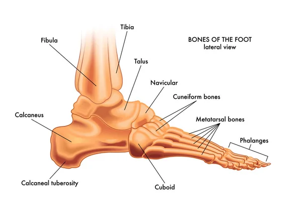
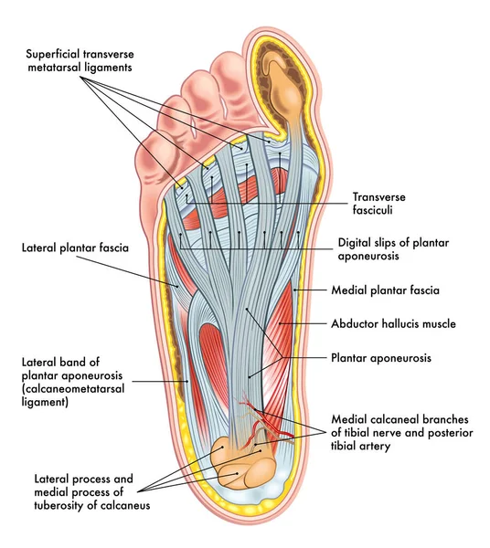
Medical Illustration Of The Major Parts Of The Foot Bones In Lateral View, With Annotations.
Vector, 5.61MB, 3072 × 2127 eps


Medical Education Chart Of Biology Human Skull Diagram Raster. Front Aspect White Background Basic Medical Education
Image, 4.08MB, 6000 × 6000 jpg

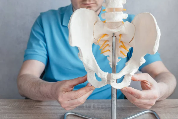
Close Up Of Male Doctor's Hand Showing Ischial Tuberosity Or Sits Bones On Skeleton Spine Model
Image, 11.16MB, 5888 × 3931 jpg
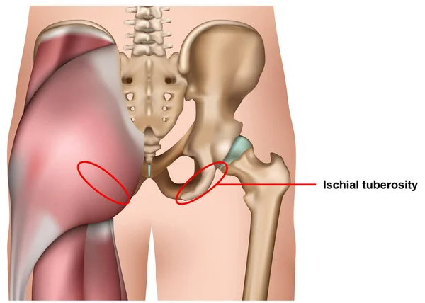
Ischial Tuberosity 3d Medical Vector Illustration Isolated On White Background Infographic
Vector, 3.29MB, 7000 × 5000 eps

Hamstring Posterior Muscle Anatomy With Bones And Ligaments Outline Diagram
Vector, 9.19MB, 3200 × 5504 eps
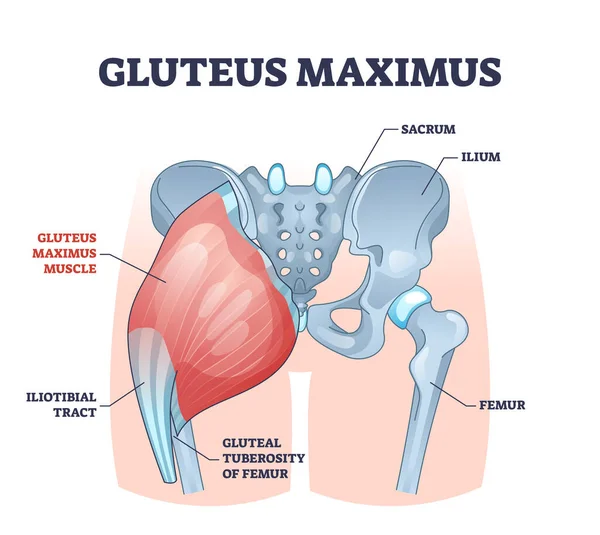
Gluteus Maximus Muscle As Medical Hip And Leg Medical Anatomy Outline Diagram
Vector, 11.49MB, 4500 × 4018 eps

Osgood-Schlatter Disease Diagram: This Illustrative Guide Visually Outlines Osgood-Schlatter Disease, A Common Knee Condition In Adolescents. It Highlights The Anatomical Aspects, Emphasizing The Inflammation At The Tibial Tuberosity.
Image, 2.12MB, 4000 × 3217 jpg
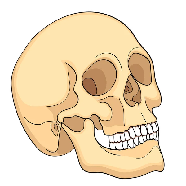
Medical Education Chart Of Biology Human Skull Diagram. Vector. Front Aspect White Background Basic Medical Education
Vector, 0.58MB, 4000 × 4000 eps
Page 1 >> Next