photo free
Home Images Vectors Videos

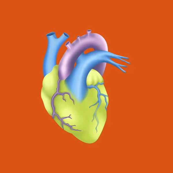
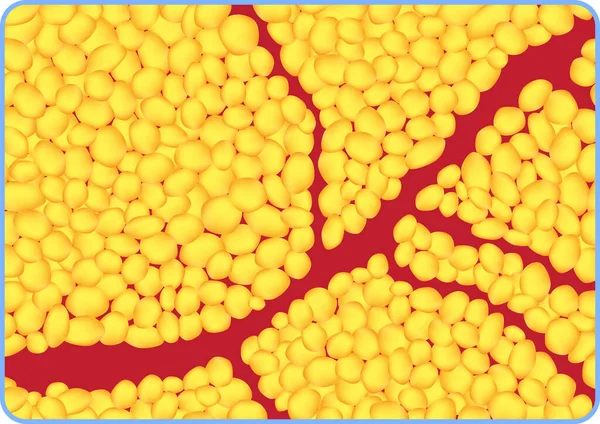
Stock image Vascularization

stock image Anatomy of the eye whose arrows represent light and revealing the lens, retina, cornea, iris, choroid. .

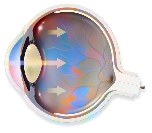
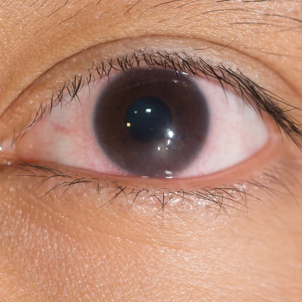
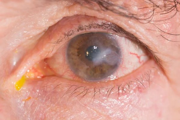
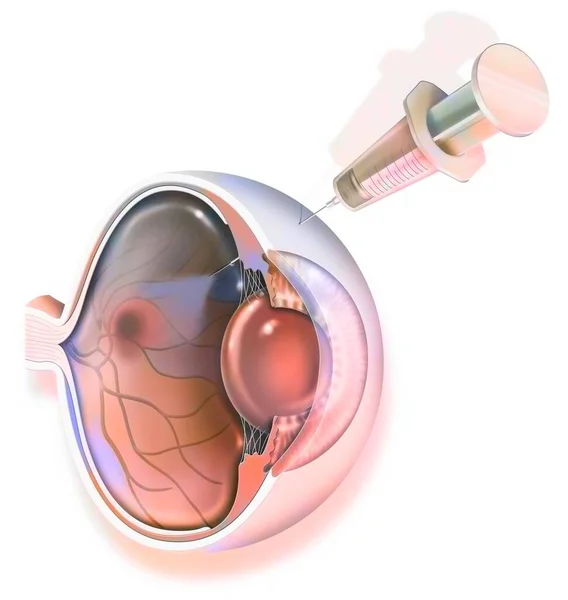
Anatomy Of The Eye Whose Arrows Represent Light And Revealing The Lens, Retina, Cornea, Iris, Choroid. .

Image, 0.76MB, 3630 × 3175 jpg
stock image EYe exam

EYe Exam

Image, 10.63MB, 6000 × 4000 jpg
stock image Liver and gall bladder with evidence of hepatic hilum.

Liver And Gall Bladder With Evidence Of Hepatic Hilum.

Image, 1.11MB, 4819 × 3780 jpg
stock image Treatment of macular degeneration by injection of RNA interference.

Treatment Of Macular Degeneration By Injection Of RNA Interference.

Image, 1.46MB, 3630 × 3500 jpg
stock image Urinary system from kidney to glomerulus with structures of kidney and ureter.

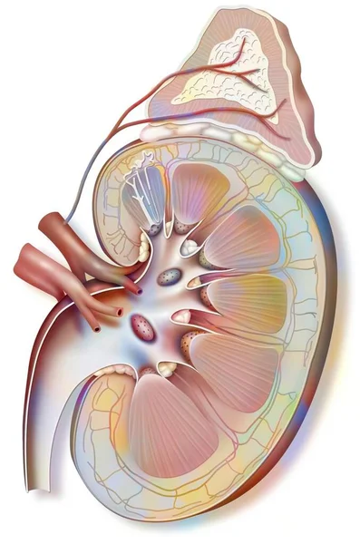
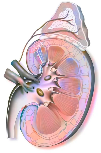
Urinary System From Kidney To Glomerulus With Structures Of Kidney And Ureter.

Image, 2.01MB, 6102 × 4134 jpg
stock image Human heart

Human Heart

Image, 0.99MB, 3930 × 3930 jpg
stock image CARDIOLOGY CONSULTATION MAN

CARDIOLOGY CONSULTATION MAN

Image, 1.1MB, 2620 × 3930 jpg
stock image Anterior and medial cerebral arteries

Anterior And Medial Cerebral Arteries

Image, 1.37MB, 3930 × 3930 jpg
stock image ANGIOLOGY CONSULTATION MAN

ANGIOLOGY CONSULTATION MAN

Image, 1.14MB, 3930 × 2620 jpg
stock image CEREBRAL ARTERIOGRAPHY macro view

CEREBRAL ARTERIOGRAPHY Macro View

Image, 1.77MB, 3630 × 2420 jpg
stock image EYe exam

EYe Exam

Image, 6.93MB, 4000 × 4000 jpg
stock image Human heart

Human Heart

Image, 1.05MB, 3930 × 3930 jpg
stock image Cerebral vasculature: arteries of the diencephalon, cerebellum and brainstem.

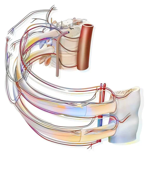
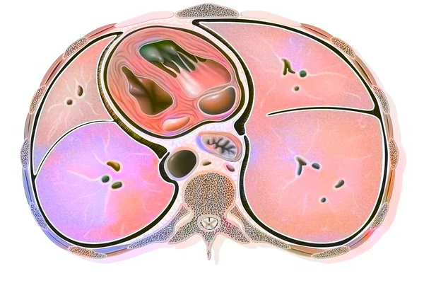
Cerebral Vasculature: Arteries Of The Diencephalon, Cerebellum And Brainstem.

Image, 1.47MB, 3630 × 3630 jpg
stock image Thorax: Heart, aorta, and pulmonary artery are visible, 3D CT scan.

Thorax: Heart, Aorta, And Pulmonary Artery Are Visible, 3D CT Scan.

Image, 11.46MB, 5003 × 4997 jpg
stock image eye exam, interstitial keratitis

Eye Exam, Interstitial Keratitis

Image, 6.26MB, 4000 × 4000 jpg
stock vector medical illustration of the formation of a subretinal neovascularization of the eye

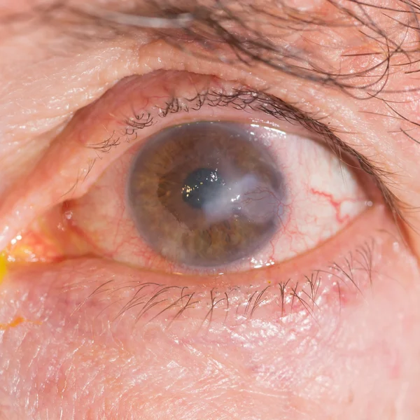
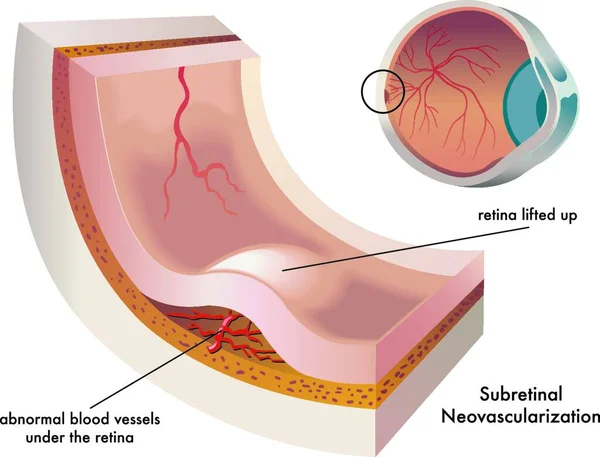
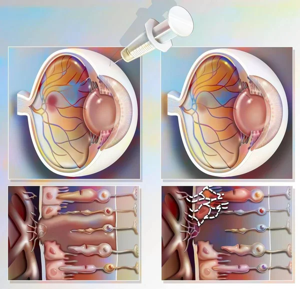
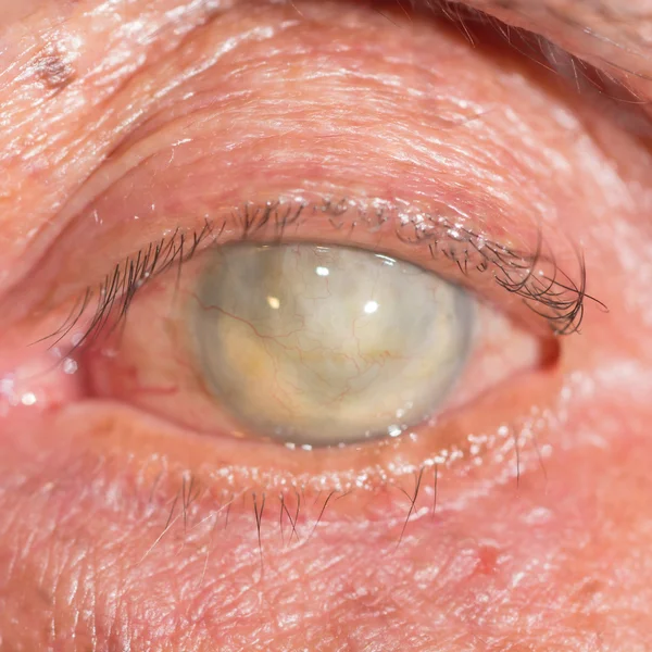
Medical Illustration Of The Formation Of A Subretinal Neovascularization Of The Eye

Vector, 0MB, 4000 × 3867 zip
stock image Brain: network of astrocytes (glial cells that support neurons).

Brain: Network Of Astrocytes (glial Cells That Support Neurons).

Image, 1.59MB, 5197 × 3352 jpg
stock image Development of bone (osteogenesis) from cartilage to adult bone.

Development Of Bone (osteogenesis) From Cartilage To Adult Bone.

Image, 1.37MB, 4978 × 3020 jpg
stock vector Histological section demonstrating yellow adipose tissue.

Histological Section Demonstrating Yellow Adipose Tissue.

Vector, 5.67MB, 10000 × 7066 ai
stock image phthisis bulbi at eye test

Phthisis Bulbi At Eye Test

Image, 6.98MB, 5563 × 3585 jpg
stock image Treatment of macular degeneration by injection of RNA interference.

Treatment Of Macular Degeneration By Injection Of RNA Interference.

Image, 0.73MB, 3630 × 3544 jpg
stock image Young woman getting massage seesion at home by a physiotherapist.

Young Woman Getting Massage Seesion At Home By A Physiotherapist.

Image, 6.95MB, 5510 × 3652 jpg
stock image Anatomy of a renal glomerulus with afferent and efferent glomerular arteriole.

Anatomy Of A Renal Glomerulus With Afferent And Efferent Glomerular Arteriole.

Image, 1.2MB, 4205 × 3661 jpg
stock image Processes in derma

Processes In Derma

Image, 1.27MB, 3930 × 3930 jpg
stock image FOOD SUPPLEMENT

FOOD SUPPLEMENT

Image, 2.17MB, 3930 × 2620 jpg
stock image eye exam, interstitial keratitis

Eye Exam, Interstitial Keratitis

Image, 8.8MB, 6000 × 4000 jpg
stock image Processes in derma

Processes In Derma

Image, 1.31MB, 3930 × 3930 jpg
stock image Left kidney (capped with its adrenal gland) and ureter.

Left Kidney (capped With Its Adrenal Gland) And Ureter.

Image, 1.47MB, 3020 × 4562 jpg
stock image phthisis bulbi at eye test

Phthisis Bulbi At Eye Test

Image, 6.12MB, 4000 × 4000 jpg
stock image Intercostal space with blood vessels and nerves.

Intercostal Space With Blood Vessels And Nerves.

Image, 1.82MB, 3937 × 4409 jpg
stock image Cross section of the thorax with the heart, lungs, esophagus and aorta.

Cross Section Of The Thorax With The Heart, Lungs, Esophagus And Aorta.

Image, 2MB, 4530 × 3020 jpg
stock image Left kidney (capped with its adrenal gland) and ureter.

Left Kidney (capped With Its Adrenal Gland) And Ureter.

Image, 1.48MB, 3020 × 4562 jpg
stock image CARDIOLOGY CONSULTATION WOMAN

CARDIOLOGY CONSULTATION WOMAN

Image, 1.36MB, 3930 × 2620 jpg
stock image EYe exam

EYe Exam

Image, 10.69MB, 4950 × 3643 jpg
stock image ANGIOLOGY CONSULTATION MAN

ANGIOLOGY CONSULTATION MAN

Image, 1.16MB, 2620 × 3930 jpg
stock image Hair in the skin with the sebaceous gland and the horripilator muscle.

Hair In The Skin With The Sebaceous Gland And The Horripilator Muscle.

Image, 1.4MB, 3020 × 4789 jpg
stock image Brain: network of astrocytes (glial cells that support neurons).

Brain: Network Of Astrocytes (glial Cells That Support Neurons).

Image, 1.6MB, 5197 × 3352 jpg
stock image Eye: treatment of macular degeneration by RNA interference.

Eye: Treatment Of Macular Degeneration By RNA Interference.

Image, 0.73MB, 3442 × 3630 jpg
stock image Processes in derma

Processes In Derma

Image, 1.31MB, 3930 × 3930 jpg
stock image ANGIOLOGY CONSULTATION MAN

ANGIOLOGY CONSULTATION MAN

Image, 1.52MB, 3930 × 2620 jpg
stock image Human urinary system

Human Urinary System

Image, 0.66MB, 3930 × 3930 jpg
stock image ANGIOLOGY CONSULTATION MAN

ANGIOLOGY CONSULTATION MAN

Image, 1.49MB, 3930 × 2620 jpg
stock image The arterial blood supply to the neck (carotids and vertebral arteries).

The Arterial Blood Supply To The Neck (carotids And Vertebral Arteries).

Image, 1.23MB, 3543 × 5057 jpg
Page 1 >> Next