
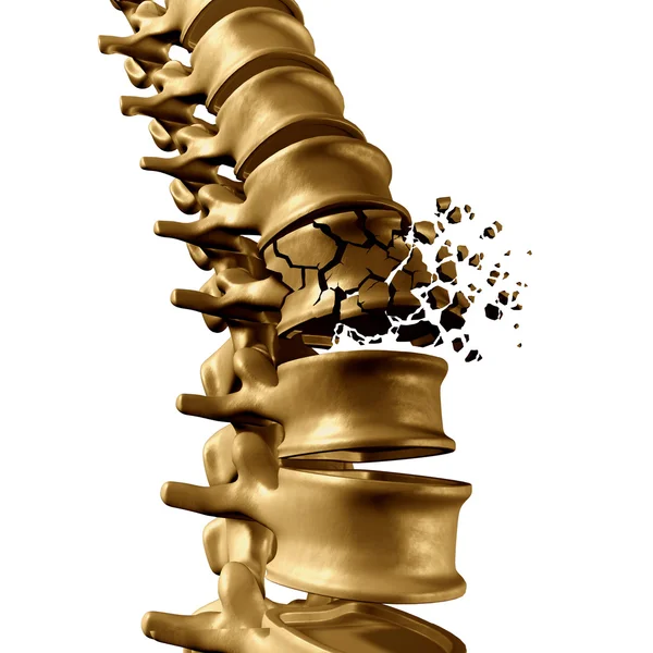
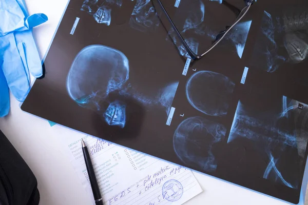
Stock image Vertebral Fracture

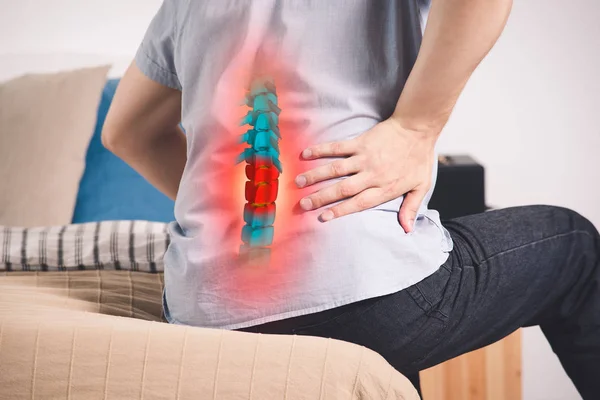
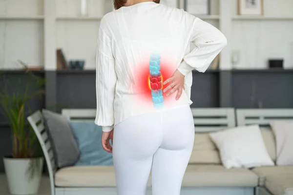
Lumbar Intervertebral Spine Hernia, Woman With Back Pain At Home, Spinal Disc Disease, Health Problems Concept
Image, 24.12MB, 7952 × 5304 jpg
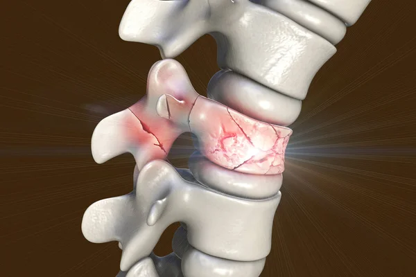
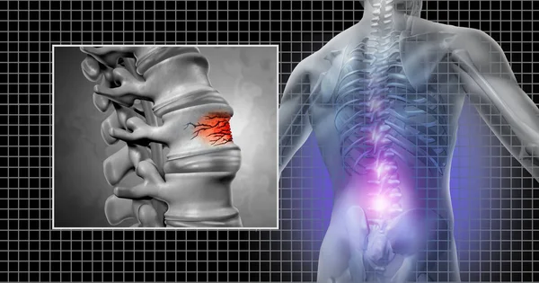
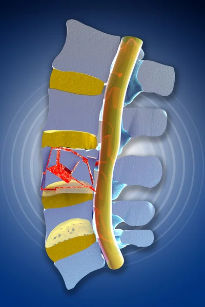
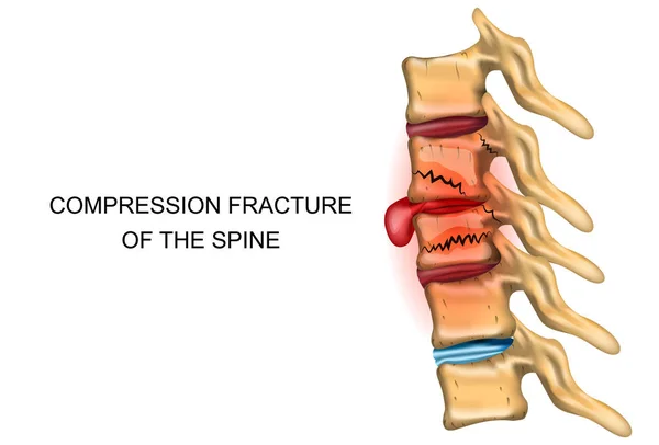
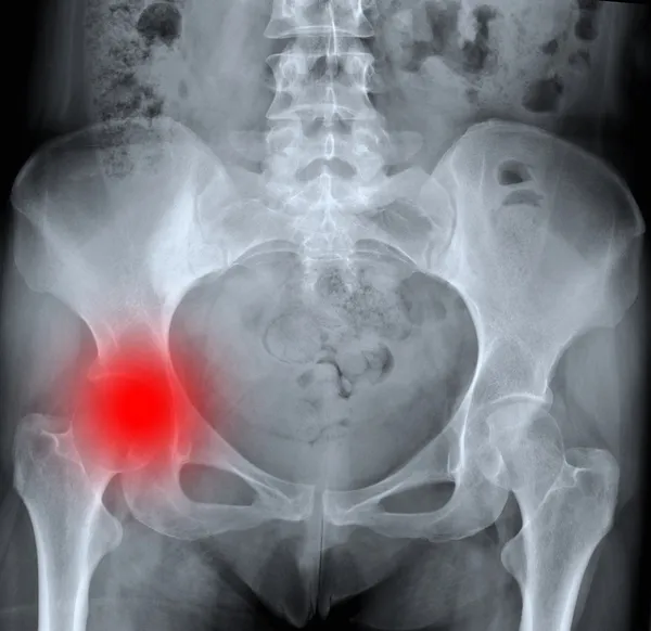
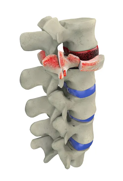
Spinal Fracture, Traumatic Vertebral Injury, 3D Illustration. Compression Fracture Of The Spine
Image, 11.05MB, 6000 × 4000 jpg

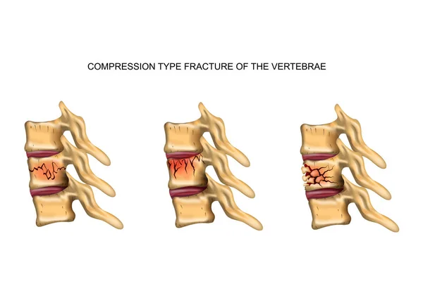
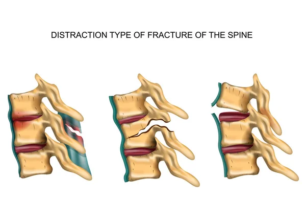
Vector Medical Illustration Of The Symptoms Of Vertebral Compression Fracture
Vector, 0MB, 3843 × 4000 zip
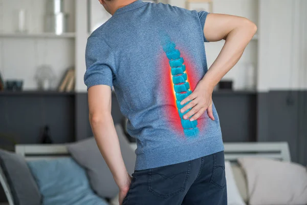
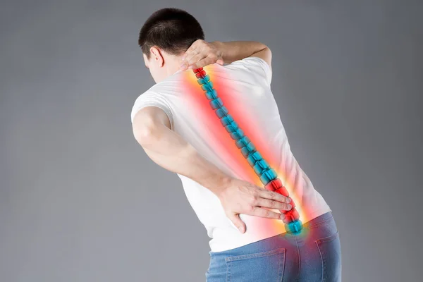
Pain In The Spine, A Man With Backache At Home, Injury In The Lower Back, Photo With Highlighted Skeleton
Image, 12.16MB, 6048 × 4032 jpg
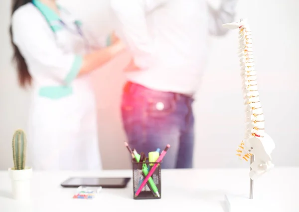
Male Patient At The Appointment With A Doctor Vertebrologist. Diagnosis And Treatment Of Back Pain Diseases, Background. Dislocation And Trauma Of The Intervertebral Disc, Pinched Nerve, Spinal Deformities
Image, 3.39MB, 5148 × 3648 jpg

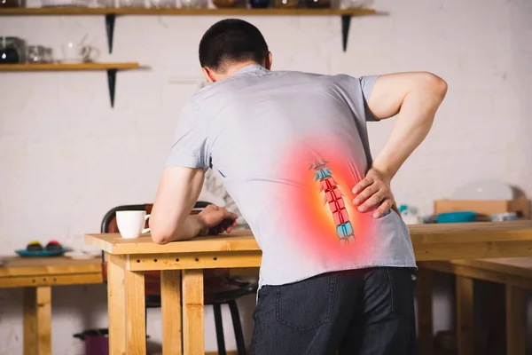
Pain In The Spine, A Man With Backache At Home, Injury In The Lower Back, Photo With Highlighted Skeleton
Image, 10.63MB, 6048 × 4032 jpg
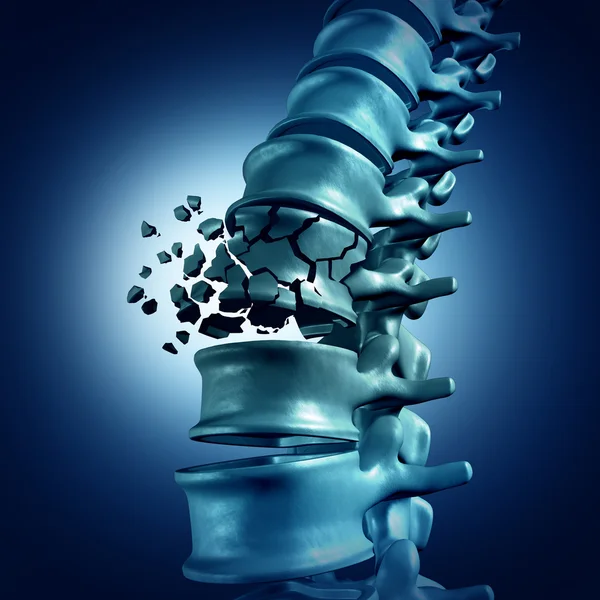
Spinal Fracture And Lower Back Pain As A Spine Injury And Vertebral Trauma As An Osteopathic Medical Concept.
Image, 6.67MB, 6662 × 3500 jpg

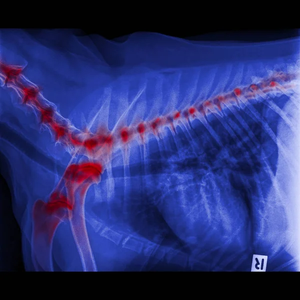
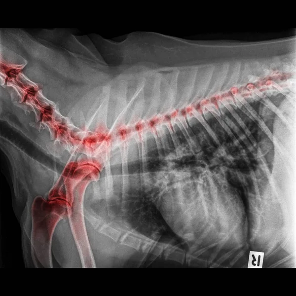
X-ray Of Dog Lateral View Closed Up Thorax And Chest Red Highlight Foreleg Bone In Shoulder Joint And Neck Bone To Back Bone- Degenerative Joint Disease In Dog- Veterinary Medicine- Veterinary Anatomy
Image, 6.73MB, 5500 × 5500 jpg
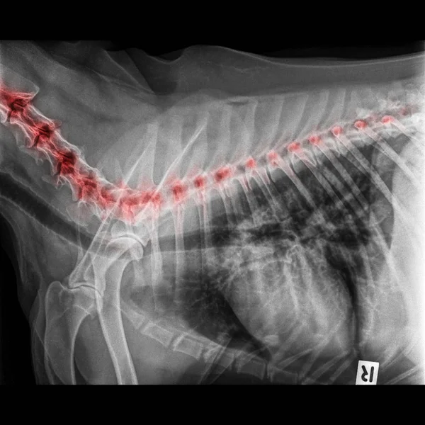
X-ray Of Dog Lateral View Closed Up Thorax And Chest Red Highlight Foreleg Bone In Shoulder Joint And Neck Bone To Back Bone- Degenerative Joint Disease In Dog- Veterinary Medicine- Veterinary Anatomy
Image, 5.39MB, 5500 × 5500 jpg
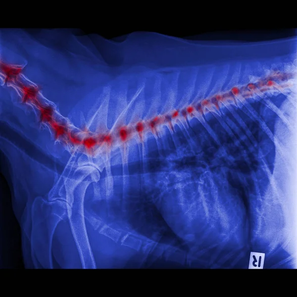
X-ray Of Dog Lateral View Closed Up In Thorax Standard And Chest With Red Highlight In Neck Bone To Back Bone Pain Areas Or Spinal Disease In Dog- Veterinary Medicine- Veterinary Anatomy Concept
Image, 6.74MB, 5500 × 5500 jpg
X-ray Of Dog Lateral View Closed Up In Thorax Standard And Chest With Red Highlight In Neck Bone To Back Bone Pain Areas Or Spinal Disease In Dog- Veterinary Medicine- Veterinary Anatomy Concept
Image, 5.26MB, 5500 × 5500 jpg
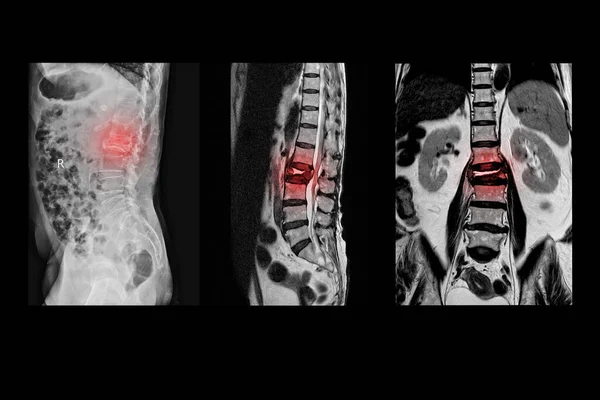
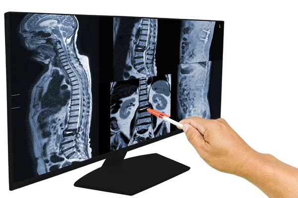
Collection MRI Of Lumbar Spine History Of Fall With Back Pain, Radiate To Leg, Rule Out Spinal Stenosis .Impression:Burst Fracture Of L2 Vertebral Body With Severe Vertebral Collapse.Medical Concept.
Image, 8.31MB, 5820 × 3890 jpg
MRI OF THORACOLUMBAR SPINE IMPRESSION: Moderate Pathological Compression Of T11 And L2 Levels With Enhancing Multiple Marrow Lesions At T1, T10 ToT12, L2, L3 To L5 Levels.
Image, 4.96MB, 5688 × 3643 jpg

X-ray Spine Lateral Views History A Male Accident And Blackpain Showing Compression Fracture Body Spine L1. Normal Disc Spaces And Paravertebral Soft Tissue.Medical Concept.
Image, 4.91MB, 6000 × 4000 jpg
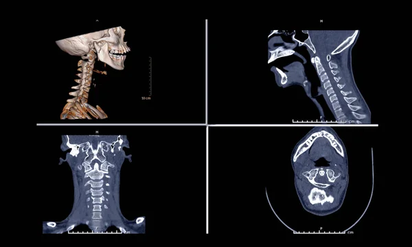
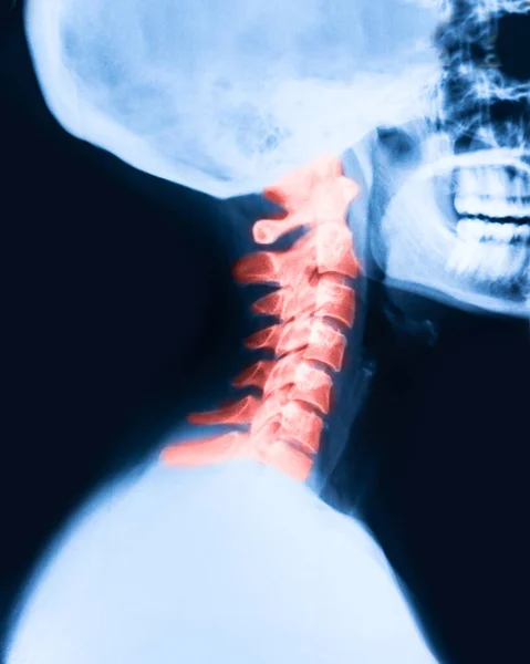
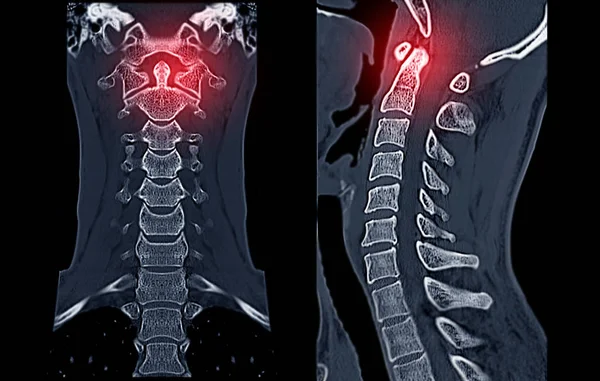
Comparison Of CT C-Spine Or Cervical Spine 3D Rendering Image , Sagittal ,Corona And Axiall View In Patient Trauma Head Injury.
Image, 2.06MB, 5008 × 3008 jpg

Vertebral Column. All Vertebrae Cervical Thoracic Lumbar Sacral And Coccygeal. Medical Science Education Human Body Anatomy Infographic
Vector, 0.5MB, 5000 × 7500 eps

MRI Of Lumbar Spine History Of Fall With Back Pain, Radiate To Leg, Rule Out Spinal Stenosis .Burst Fracture Of L2 Vertebral Body With Severe Vertebral Collapse.
Image, 4.67MB, 6000 × 4000 jpg
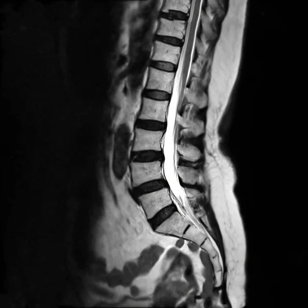
MRI L-s Spine Or MRI Of Lambosacral Spine Or L-S Spine On Sagittal Plane T2 Technique For Diagnosis Spinal Cord Compression.
Image, 2.02MB, 3072 × 3072 jpg

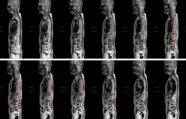
MRI OF THORACOLUMBAR SPINE:IMPRESSION: Moderate Pathological Compression Of T11 And L2 Levels With Enhancing Multiple Marrow Lesions At T1, T10 ToT12, L2, L3 To L5 Levels.multiple Bone Metastases Should Be Considered,hematologic Malignancy .
Image, 7.76MB, 5760 × 4348 jpg

Lumbar Intervertebral Spine Hernia, Man With Back Pain At Home, Spinal Disc Disease, Painful Area Highlighted In Red
Image, 25.11MB, 7952 × 5304 jpg
Lumbar Intervertebral Spine Hernia, Woman With Back Pain At Home, Spinal Disc Disease, Health Problems Concept
Image, 19.24MB, 7952 × 5304 jpg

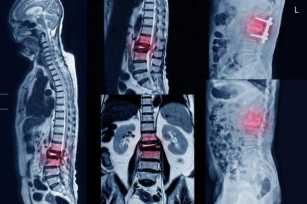
The Doctor Reported The MRI Scans Of The Lumbar Spine Compression Fracture Bulging Of L1-L2. And Post Operation Fixed By Iron Rod And Screws. Medical Education Concept.
Image, 7.51MB, 6000 × 4000 jpg

Human Spine Anatomy Vector Illustration.Spine Medical Center, Clinic, Institute, Diagnostic Element. Spinal Icon
Vector, 3.62MB, 5000 × 5000 eps
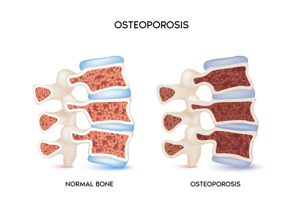
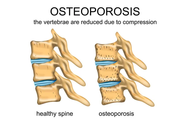
Spinal Bones, Healthy And Unhealthy Bone, Osteoporosis. Medical Or Healthcare Concept. Bone Protection. Isolated On A White Background. Realistic 3d Vector Illustration
Vector, 3.58MB, 5208 × 3753 eps
Pain In The Spine, A Man With Backache, Injury In The Human Back, Chiropractic Treatments Concept
Image, 11.76MB, 6048 × 4032 jpg
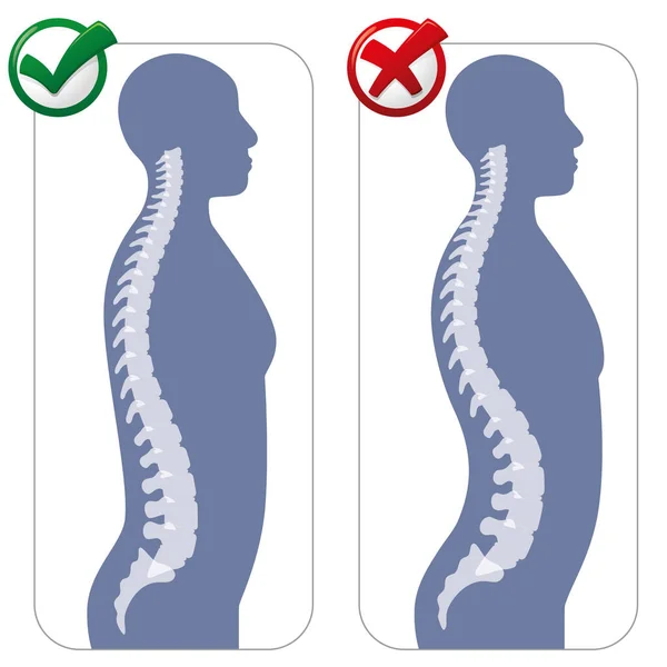
Human Anatomy Lumbar Hyper Lordosis Illustration. Ideal For Catalogues, Newsletters And Medical Guides
Vector, 2.7MB, 5000 × 5000 eps

Concept Of Backaches, Back Pain Or Manifestations Or Symptoms Of Radiculopathy. Man Grabbed With Palm Over Lumbar Region On Back, Indicating Localization Of Pain In Spine Pathology
Image, 12.69MB, 6000 × 4000 jpg
Page 1 >> Next