
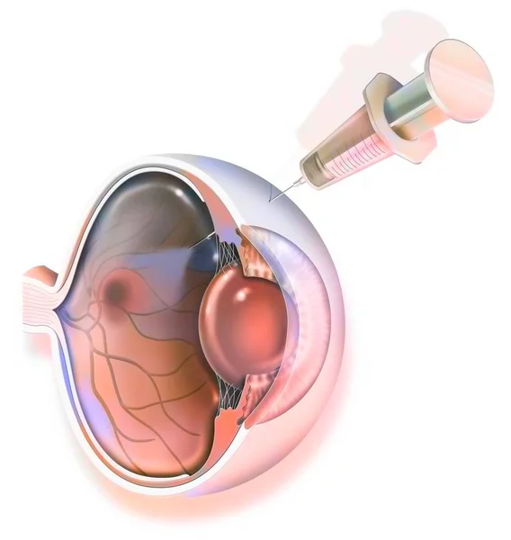
Stock image Vitreous Humor
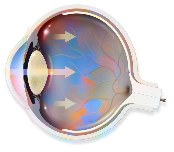
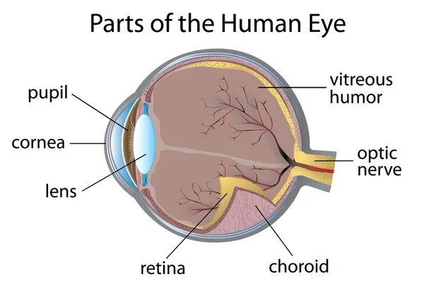
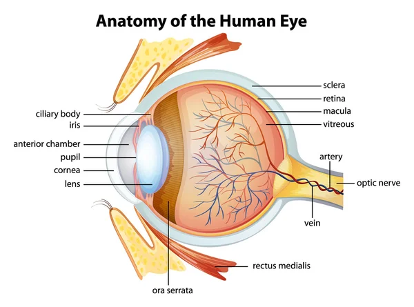
Anatomy Of The Eye Whose Arrows Represent Light And Revealing The Lens, Retina, Cornea, Iris, Choroid. .
Image, 0.76MB, 3630 × 3175 jpg


Retinal Detachment. Cross Section Of A Normal Human Eyes And Disorder Of The Eye In Which The Retina Peels. Schematic Diagram. Detailed Vector Illustration
Vector, 4.17MB, 6309 × 3000 eps


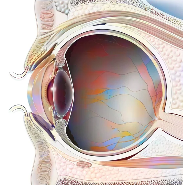
The Posterior Chamber, Trabecular Meshwork, And Eye Drainage. Crucial Role In Removing Intraocular Fluid. Intraocular Pressure. Eye Health Concept, Glaucoma Treatment Research Vector Illustration
Vector, 0.39MB, 5787 × 3916 eps

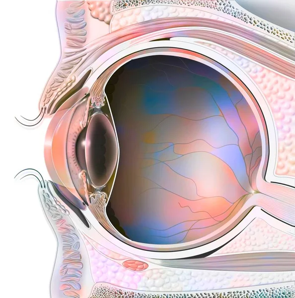
The Posterior Chamber, Trabecular Meshwork, And Eye Drainage. Crucial Role In Removing Intraocular Fluid. Intraocular Pressure. Eye Health Concept, Glaucoma Treatment Research Vector Illustration
Vector, 0.4MB, 4916 × 4064 eps

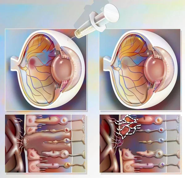
The Posterior Chamber, Trabecular Meshwork, And Eye Drainage. Crucial Role In Removing Intraocular Fluid. Intraocular Pressure. Eye Health Concept, Glaucoma Treatment Research Vector Illustration
Vector, 0.43MB, 4774 × 3946 eps

The Posterior Chamber, Trabecular Meshwork, And Eye Drainage. Crucial Role In Removing Intraocular Fluid. Intraocular Pressure. Eye Health Concept, Glaucoma Treatment Research Vector Illustration
Vector, 0.43MB, 4774 × 3946 eps

The Posterior Chamber, Trabecular Meshwork, And Eye Drainage. Crucial Role In Removing Intraocular Fluid. Intraocular Pressure. Eye Health Concept, Glaucoma Treatment Research Vector Illustration
Vector, 0.48MB, 7897 × 2624 eps

The Posterior Chamber, Trabecular Meshwork, And Eye Drainage. Crucial Role In Removing Intraocular Fluid. Intraocular Pressure. Eye Health Concept, Glaucoma Treatment Research Vector Illustration
Vector, 0.48MB, 7897 × 2624 eps

The Posterior Chamber, Trabecular Meshwork, And Eye Drainage. Crucial Role In Removing Intraocular Fluid. Intraocular Pressure. Eye Health Concept, Glaucoma Treatment Research Vector Illustration
Vector, 0.4MB, 5467 × 4014 eps

The Posterior Chamber, Trabecular Meshwork, And Eye Drainage. Crucial Role In Removing Intraocular Fluid. Intraocular Pressure. Eye Health Concept, Glaucoma Treatment Research Vector Illustration
Vector, 0.4MB, 4917 × 4064 eps

A Medical Illustration Depicting Vitreous Hemorrhage Observed During Ophthalmoscopy, Revealing Blood Within The Vitreous Humor Obscuring Retinal Details.
Image, 2.99MB, 5000 × 5000 jpg

The Posterior Chamber, Trabecular Meshwork, And Eye Drainage. Crucial Role In Removing Intraocular Fluid. Intraocular Pressure. Eye Health Concept, Glaucoma Treatment Research Vector Illustration
Vector, 0.4MB, 4916 × 4064 eps
The Posterior Chamber, Trabecular Meshwork, And Eye Drainage. Crucial Role In Removing Intraocular Fluid. Intraocular Pressure. Eye Health Concept, Glaucoma Treatment Research Vector Illustration
Vector, 0.33MB, 5787 × 3916 eps

The Posterior Chamber, Trabecular Meshwork, And Eye Drainage. Crucial Role In Removing Intraocular Fluid. Intraocular Pressure. Eye Health Concept, Glaucoma Treatment Research Vector Illustration
Vector, 0.44MB, 8009 × 3022 eps
Page 1 >> Next