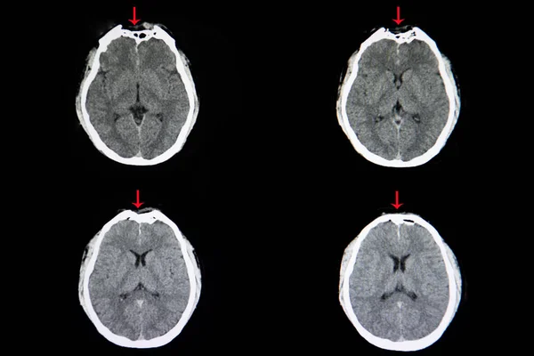
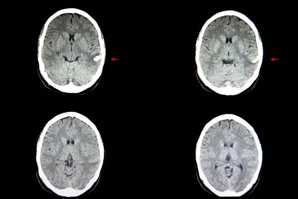
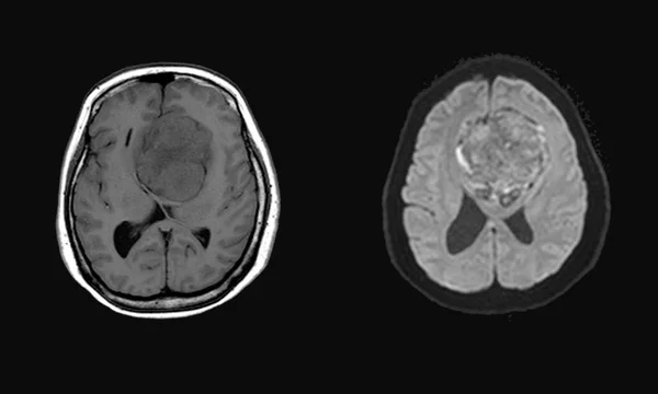
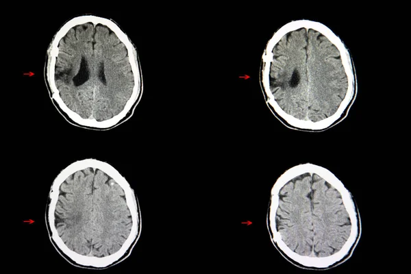
Stock image A CT brain scan show a metastatic cancer masses in the right thalamic and basal ganglionic areas with some blood in the tumor masses and the right ventricle.

Published: Jul.15, 2020 07:21:43
Author: navuths@gmail.com
Views: 3
Downloads: 0
File type: image / jpg
File size: 7.18 MB
Orginal size: 6240 x 4160 px
Available sizes:
Level: beginner