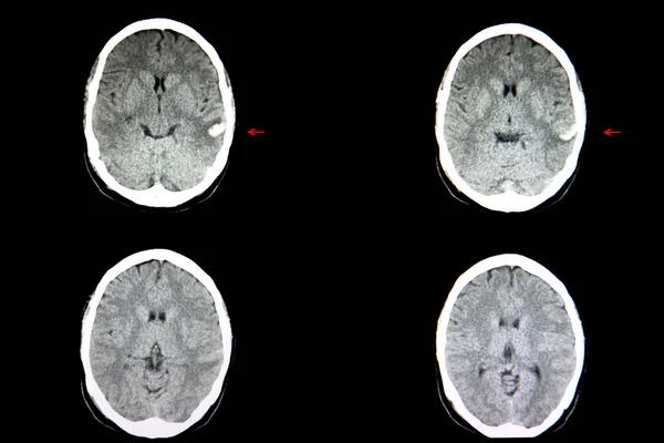
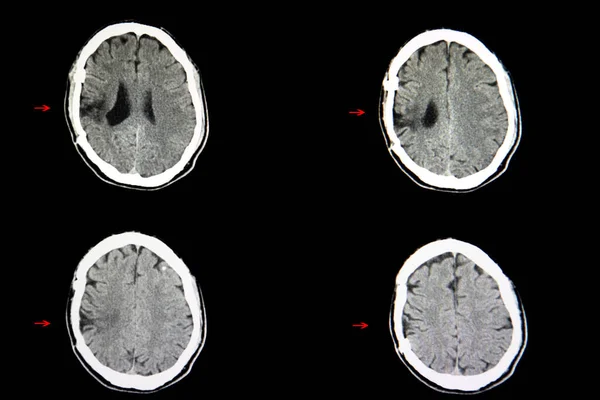
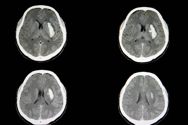
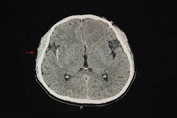
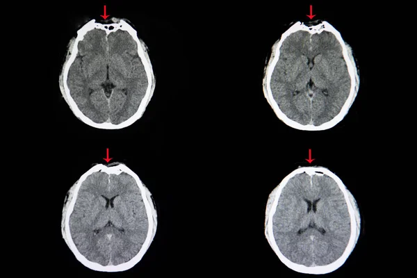
Stock image CT scan image of skull depression fracture at frontal area.
Published: Jul.15, 2020 11:58:43
Author: navuths@gmail.com
Views: 4
Downloads: 0
File type: image / jpg
File size: 9.59 MB
Orginal size: 6240 x 4160 px
Available sizes:
Level: beginner