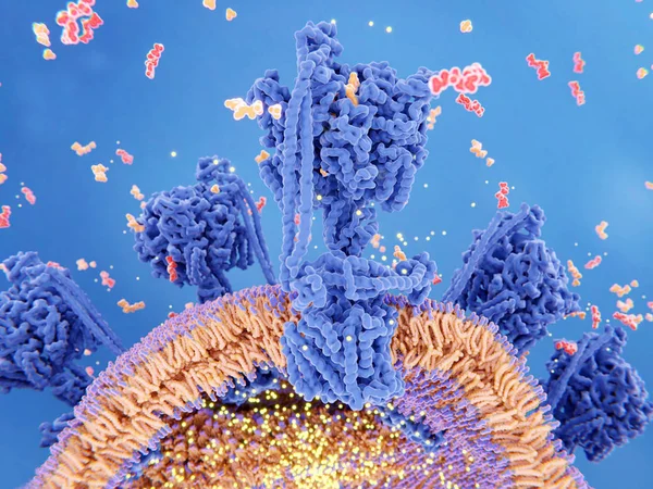
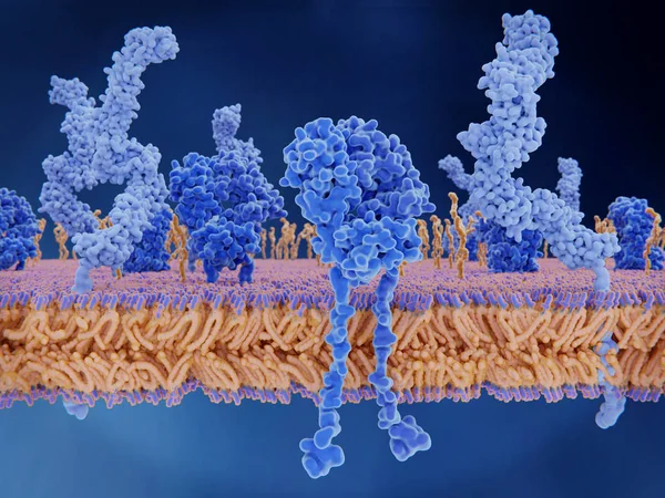
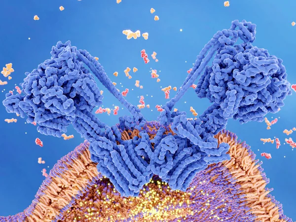
Stock image ATP synthase couples ATP (red) synthesis from ADP and inorganic phosphate (orange) to a proton gradient (yellow) created across the mitochondrial membrane during cellular respiration.
Published: Mar.15, 2019 08:15:18
Author: animaxx3d
Views: 118
Downloads: 13
File type: image / jpg
File size: 12.41 MB
Orginal size: 8000 x 6000 px
Available sizes:
Level: bronze