
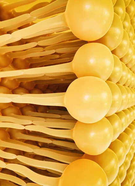
Stock image Lipid Bilayer

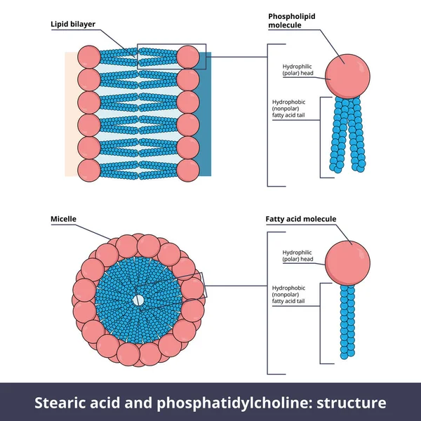
Structure Of Two Lipids. Stearic Acid (fatty Acid) And Phosphatidylcholine (phospholipid) Are Composed Of Chemical Groups That Form Polar Heads (hydrophilic) And Nonpolar Tails" (hydrophobic).
Vector, 10.84MB, 5208 × 5208 eps
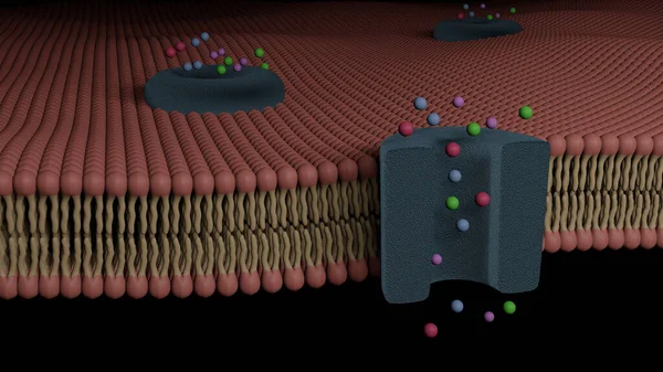
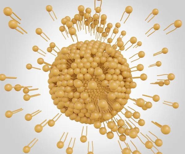
Glycoprotein Bilayer Cell Membrane Cross Section And Its Ion Channel (3D Rendering)
Image, 18.38MB, 8000 × 4500 jpg


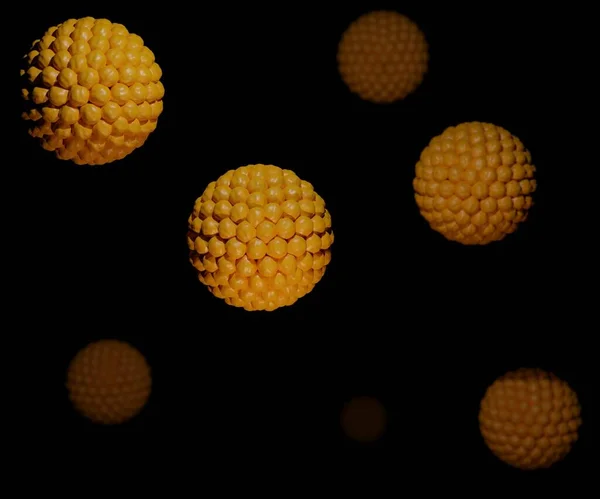
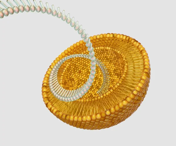
Lipid Bilayer-coated Mesoporous Silica Nanoparticles: Emerging Nanocarriers For Targeted Drug Delivery And 3D-rendered Nanomedicine Release
Image, 0.81MB, 3840 × 2160 jpg
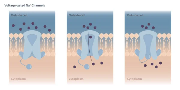
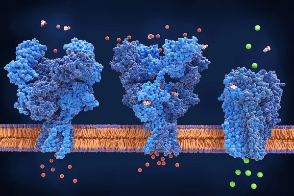
Neuronal Charged Membranes. Voltage-gated Ion Channels Are Closed At The Resting Potential And Open In Response To Changes In Membrane Voltage.
Vector, 7.67MB, 8334 × 4167 eps


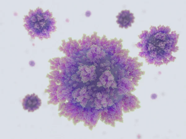
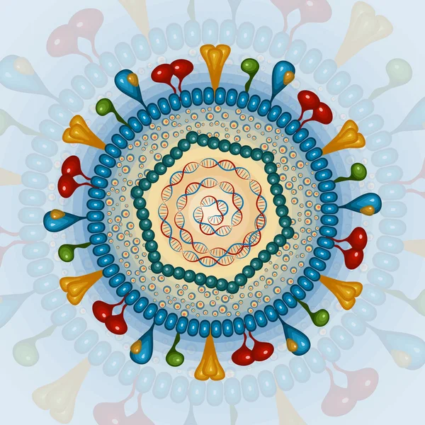
Coronavirues, COVID-19. The SARS-CoV-2 Virus Has 3 Surface Proteins. Attached To A Lipid Bilayer. The Biggest Is The Spike (S) Protein. Source: PDB Entries 6vsb, 5x29.
Image, 6.31MB, 8000 × 6000 jpg
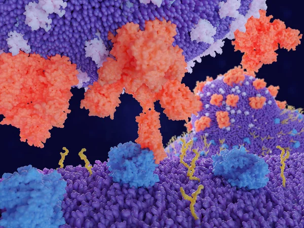
Binding Of The Coronavirus Spike Protein(red) To An ACE2 Receptor (blue) On A Human Cell Leads To The Penetration Of The Virus In The Cell, As Depicted In The Background.
Image, 8.86MB, 8000 × 6000 jpg

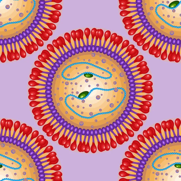
Transferosomes Structure Are Vesicular Carrier Systems That Enclosed By A Lipid Bilayer, Together With An Edge Activator 3d Rendering
Image, 0.53MB, 2400 × 2000 jpg

A 3D Rendering Featuring A Half-cut Liposome And Other Liposomes Dispersed On A White Background
Image, 0.6MB, 3840 × 2160 jpg

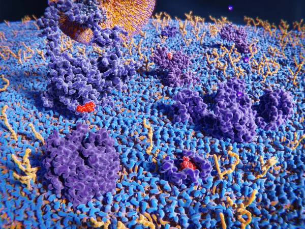
Membrane Proteins (violett), Glycolipids (yellow) And Several Ligands Of The Proteins
Image, 7.89MB, 8000 × 6000 jpg
The Liposomes Can Burst Or Be Broken Down To Release Nanodrugs Or Nanomedicine 3d Rendering
Image, 0.36MB, 2400 × 2000 jpg


3d Rendering Of Liposomes Within Liposomes Are Known As Multivesicular Liposomes Or Nested Liposomes.
Image, 0.58MB, 3840 × 2160 jpg

Coronaviruses Penetrating In Human Cell. Binding Of The Coronavirus Spike Protein(red) To An ACE2 Receptor (blue) Leads To The Penetration Of The Virus In The Cell. 3d Rendering. PDB 6VSB, 6ACJ
Image, 11.34MB, 8000 × 6000 jpg

The Liposomes Can Burst Or Be Broken Down To Release Nanodrugs Or Nanomedicine 3d Rendering
Image, 0.61MB, 2400 × 2000 jpg

3d Rendering Of Liposomes Within Liposomes Are Known As Multivesicular Liposomes Or Nested Liposomes.
Image, 0.92MB, 3840 × 2160 jpg
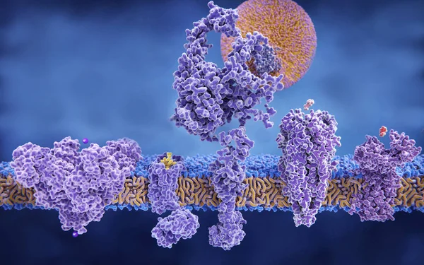
5 Membrane Proteins With Their Ligands: (left To Right) Potassium Channel, Delta-opioid Receptor, LDL Receptor, Acetylcholine Receptor, Histamine Receptor.
Image, 4.48MB, 8000 × 5000 jpg

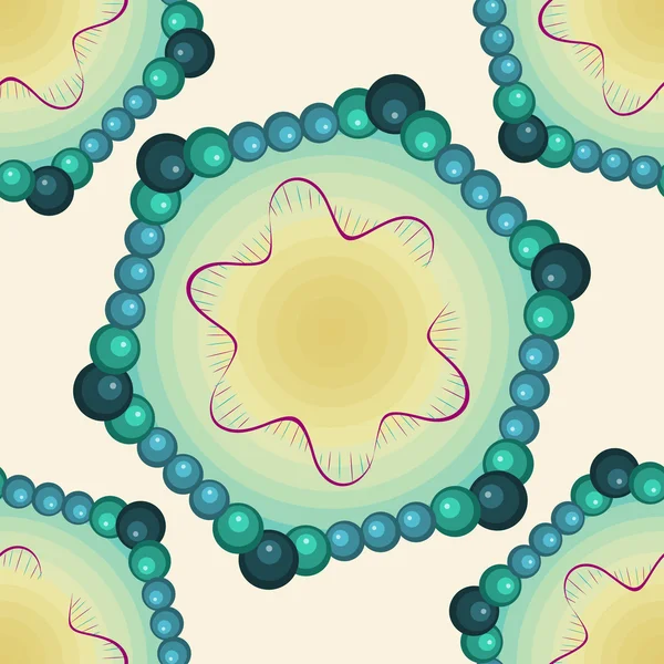
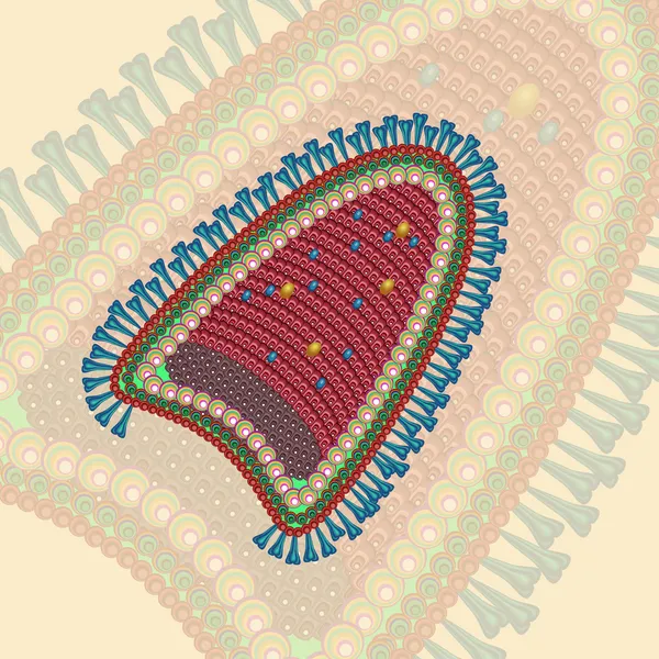
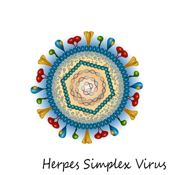
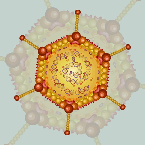
Structure And Anatomy Of Virions. Difference Between Naked Nucleocapsid Virus And Enveloped Virus. Vector Illustration
Vector, 3.38MB, 5000 × 3785 eps

A 3D Rendering Featuring A Half-cut Liposome And Other Liposomes Dispersed On A White Background
Image, 0.48MB, 3840 × 2160 jpg

3d Rendering Of Liposomes Within Liposomes Are Known As Multivesicular Liposomes Or Nested Liposomes.
Image, 0.81MB, 3840 × 2160 jpg


Antibody Binding To The Spike Protein Of The SARS-CoV-2 Virus Is An Essential Step For Developing Immunity To The Coronavirus. PDB Source 6VSB, 1IGT
Image, 6.16MB, 8000 × 6000 jpg
A Cell Membrane In Fish Eye Perspective. Receptors: Opioid Receptor, LDL Receptor And Acetyl Choline Receptor. Channel Proteins: Chloride Channel And Acetat Permease. The Glycolipids Are Depicted In Bluish Green.
Image, 15.26MB, 8000 × 6000 jpg
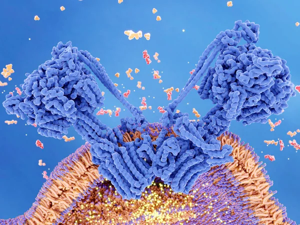
ATP Synthase Couples ATP (red) Synthesis From ADP And Inorganic Phosphate (orange) To A Proton Gradient (yellow) Created Across The Mitochondrial Membrane During Cellular Respiration.
Image, 12.41MB, 8000 × 6000 jpg
Liposomes As Nanocarriers For Small Interference RNA (siRNA) Delivery 3d Rendering
Image, 0.2MB, 2400 × 2000 jpg
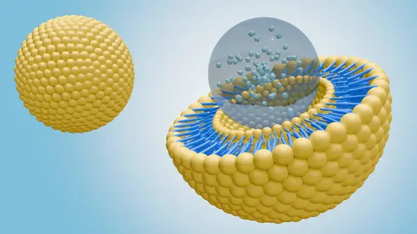

A 3D Rendering Featuring A Half-cut Liposome And Other Liposomes Dispersed On A White Background
Image, 0.54MB, 3840 × 2160 jpg

3d Rendering Of Liposomes Within Liposomes Are Known As Multivesicular Liposomes Or Nested Liposomes.
Image, 0.61MB, 3840 × 2160 jpg

From Bottom To Top: Fo-subunit (violet), Axle-element (light Violet), The Stator Element (light Blue), And ATP Synthesis Takes Place In The Upper F1-subunit (blue).
Image, 2.27MB, 8000 × 6000 jpg
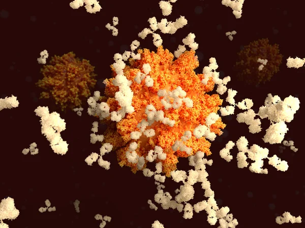
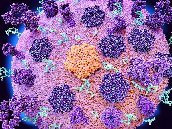
Antibodies Binding To A Coronavirus. Binding Of Antibodies To The Spike (S)-protein Of The SARS-CoV-2 Virus Is An Essential Step For Developing Immunity To The Coronavirus.
Image, 4.3MB, 8000 × 6000 jpg

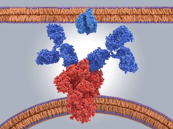
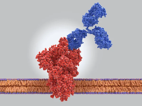
Antibodies Binding To The Spike Protein (red) Of The Coronavirus Prevent It's Binding To ACE2 (light Blue) On A Human Cell.
Image, 6.75MB, 8000 × 6000 jpg

Page 1 >> Next