Home
Images
Vectors
Videos
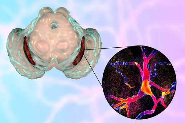
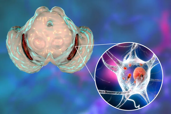
Stock image Black substance of the midbrain and its dopaminergic neurons, 3D illustration. Black substance regulates movement and reward, its degeneration is a key step in development of Parkinson's disease
Published:
Jan.28, 2021 09:43:21
Author:
katerynakon
Views:
15
Downloads:
1
File type:
image
/
jpg
File size:
2.54 MB
Orginal size:
6000
x
4000
px
Available sizes:
S
(500 x 333)
M
(1000 x 667)
L
(2000 x 1333)
XL
(6000 x 4000)
EL0
(6000 x 4000)
Level:
silver
Download Image
Image topics:
Close Up
Macro
Illustration
Isolated
Model
Medicine
Healthcare
Medical
3d
Concept
Cell
Development
Disease
System
Conceptual
Destruction
Nucleus
Brain
Neuron
Nervous
Disorder
Neurology
Microscopic
Inflammation
Dementia
Progressive
Schizophrenia
Degeneration
Neuronal
Neurone
Neurological
Parkinson
White Background
Nervous System
Nerve Cell
Neurodegenerative
Neurodegeneration
Parkinsons Disease
Multiple System Atrophy
Substantia Nigra
Dopaminergic
Neuronal Degeneration
Wooden Chest Syndrome
Pars Compacta
Similar stock images
Black Substance, Basal Banglia Of The Midbrain, In Parkinson's Disease, 3D Illustration Showing Decrease Of Its Volume And Degeneration Of Dopaminergic Neurons In The Pars Compacta Of The Black Substance
6000 × 4000
Black Substance Of The Midbrain And Its Dopaminergic Neurons, 3D Illustration. Black Substance Regulates Movement And Reward, Its Degeneration Is A Key Step In Development Of Parkinson's Disease
6000 × 4000
Substantia Nigra Of The Midbrain And Its Dopaminergic Neurons, 3D Illustration. Substantia Nigra Regulates Movement And Reward, Its Degeneration Is A Key Step In Development Of Parkinson's Disease
6000 × 4000
Black Substance, Basal Banglia Of The Midbrain, In Parkinson's Disease, 3D Illustration Showing Decrease Of Its Volume And Levy Bodies Inclusions In Neurons In The Pars Compacta Of Black Substance
6000 × 4000
Dorsal Striatum Highlighted In Human Brain And Close-up View Of Degrading Neurons Of Dorsal Striatum Seen In Huntington's Disease, 3D Illustration
8672 × 4878
Human Brain With Highlighted Temporal Lobe And Close-up View Of Pyramidal Neurons Found In Temporal Cortex, 3D Illustration
7802 × 4388