photo free
Home Images Vectors Videos

Stock image Comparison MRI brain Axial T1 and T2 for detect a variety of conditions of the brain such as cysts, tumors, bleeding, swelling, developmental and structural abnormalities, infections.
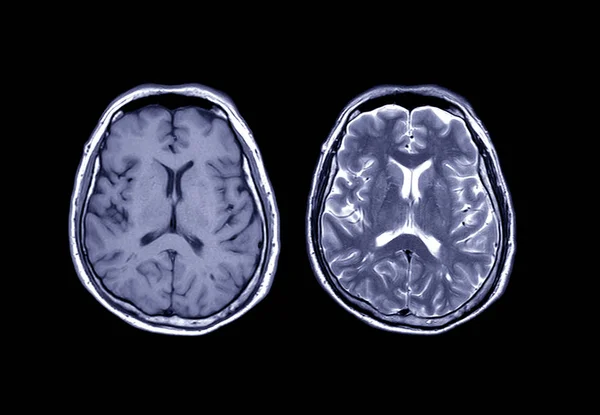
Stock image Comparison MRI brain Axial T1 and T2  for detect a variety of conditions of the brain such as cysts, tumors, bleeding, swelling, developmental and structural abnormalities, infections.
Published: Dec.23, 2019 11:47:31
Author: samunella
Views: 111
Downloads: 5
File type: image / jpg
File size: 2.45 MB
Orginal size: 4356 x 3016 px
Available sizes:
S
(500 x 346)
M
(1000 x 692)
L
(2000 x 1385)
XL
(4356 x 3016)
EL0
(4356 x 3016)
Level: beginner
Download Image
Image topics:

Similar stock images

stock image magnetic resonance image, mri scan of the brain

Magnetic Resonance Image, Mri Scan Of The Brain

6134 × 4200
stock image X-ray image of the brain computed tomography

X-ray Image Of The Brain Computed Tomography

6016 × 4000
stock image Horizontal section of a human brain

Horizontal Section Of A Human Brain

3721 × 5582
stock image Compare MRI brain  Axial T2W flair and T2W view  for showing enchepalomalacia disease.

Compare MRI Brain Axial T2W Flair And T2W View For Showing Enchepalomalacia Disease.

7194 × 4272
stock image CT scan of brain : show hemorrhagic stroke

CT Scan Of Brain : Show Hemorrhagic Stroke

3456 × 3456
stock image A CT scan of the brain of a patient with cerebral atrophy showing large ventricles and generalzed shrinkage of the cerebral tissue.

A CT Scan Of The Brain Of A Patient With Cerebral Atrophy Showing Large Ventricles And Generalzed Shrinkage Of The Cerebral Tissue.

6240 × 4160
stock image X-Ray or MRI brain scan background, magnetic resonance  tomography image

X-Ray Or MRI Brain Scan Background, Magnetic Resonance Tomography Image

3001 × 2001
stock image X-ray

X-ray

3336 × 3336