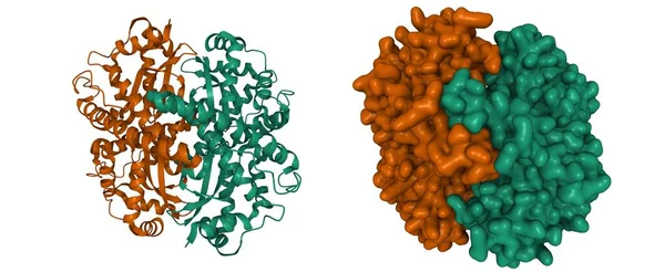
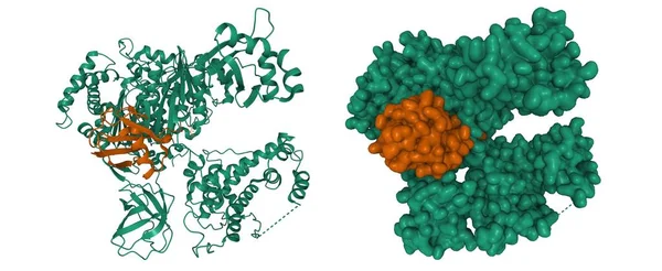
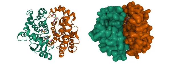
Stock image Crystal structure of human glutathione transferase (GST) A1-1 in complex with glutathione, 3D cartoon and Gaussian surface models, chain id color scheme, based on PDB 1pkw, white background
Published: Jul.19, 2021 07:26:14
Author: unnaugan
Views: 0
Downloads: 0
File type: image / jpg
File size: 5.49 MB
Orginal size: 10000 x 4000 px
Available sizes:
Level: beginner