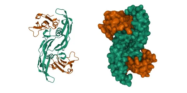
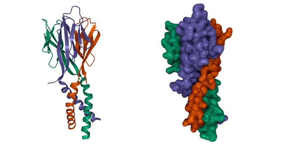
Stock image Structure of immune receptor HLA-DRB1 with vimentin bound, 3D cartoon and Gaussian surface models, chain id color scheme, based on PDB 4mdj, white background
Published: Jul.19, 2021 07:26:14
Author: unnaugan
Views: 15
Downloads: 2
File type: image / jpg
File size: 4.51 MB
Orginal size: 10000 x 4000 px
Available sizes:
Level: beginner