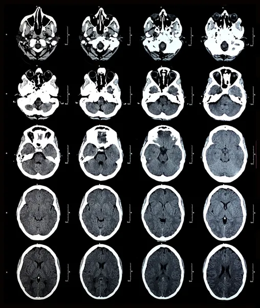
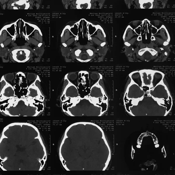
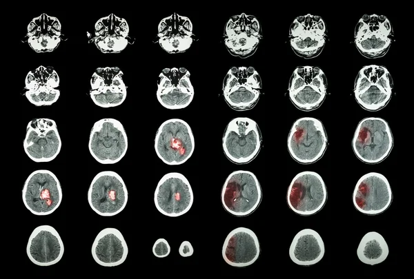
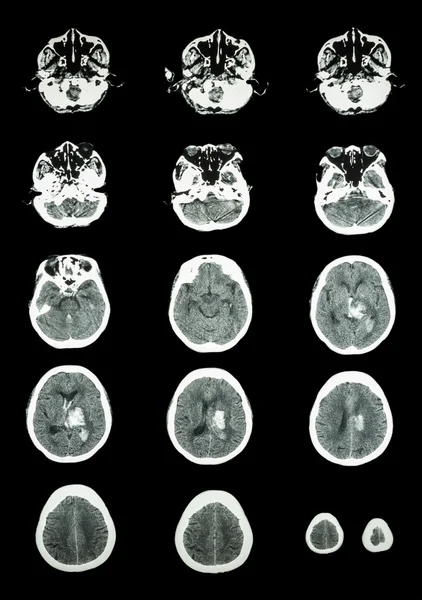
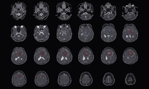
Stock image CT Scan Brain Axial scans with 5 mm slice thickness from OM-line to vertex

Published: Oct.03, 2019 14:26:59
Author: Richmanphoto
Views: 49
Downloads: 2
File type: image / jpg
File size: 1.89 MB
Orginal size: 4000 x 2430 px
Available sizes:
Level: bronze