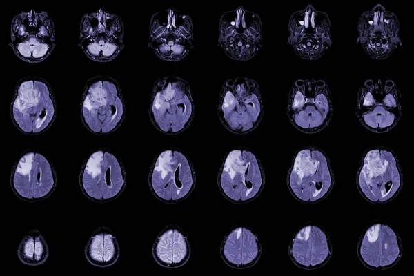
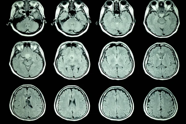
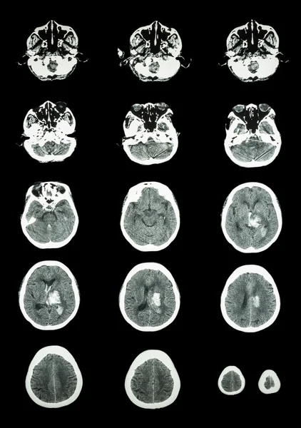
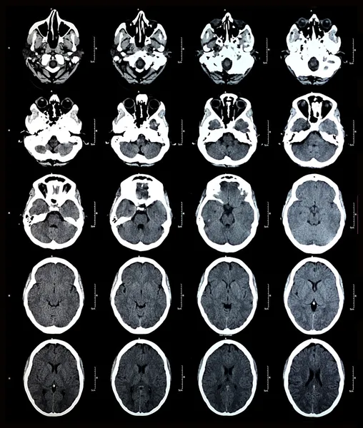
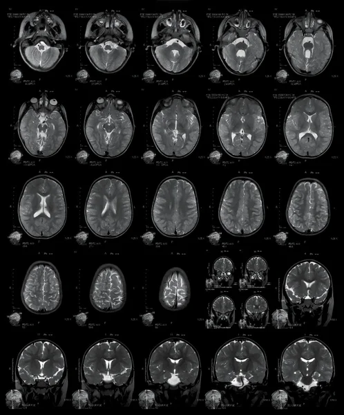
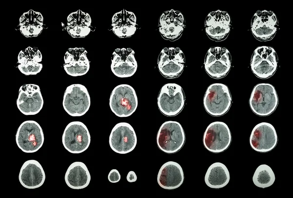
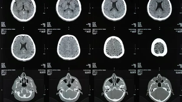
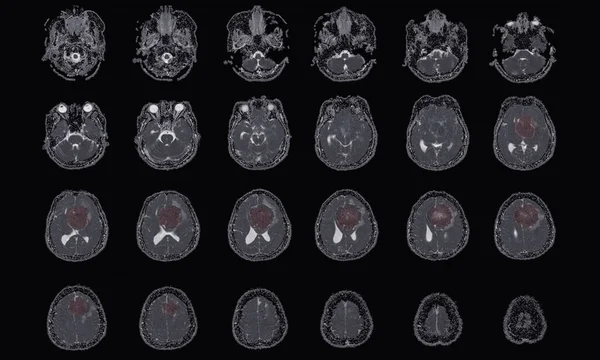
Stock image MRI BRAIN Finding of meningioma arising from anterior falx cerebri, extending to bilateral frontal regions, with adjacent minimal perilesional edema at the left frontal lobes, Medical image concept.
Published: Nov.08, 2022 16:53:53
Author: Richmanphoto
Views: 5
Downloads: 0
File type: image / jpg
File size: 6.13 MB
Orginal size: 7000 x 4205 px
Available sizes:
Level: bronze