Home
Images
Vectors
Videos
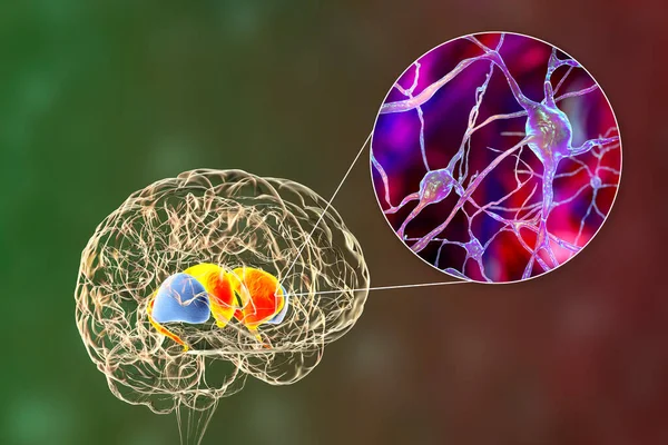
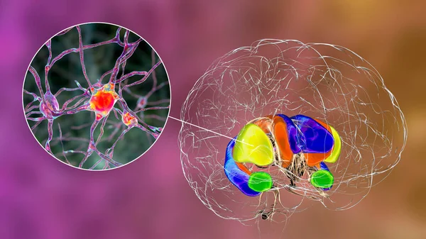
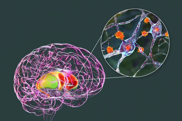
Stock image Destruction of neurons of the caudate nucleus, conceptual 3D illustration. Caudate nucleus belongs to the brain basal ganglia, its neurons are damaged in Huntingon's disease and other choreas
Published:
May.13, 2021 13:16:47
Author:
katerynakon
Views:
2
Downloads:
1
File type:
image
/
jpg
File size:
16.49 MB
Orginal size:
7996
x
5331
px
Available sizes:
S
(500 x 333)
M
(1000 x 667)
L
(2000 x 1333)
XL
(7996 x 5331)
EL0
(7996 x 5331)
Level:
silver
Download Image
Image topics:
Illustration
Human
Medicine
Medical
3d
Cell
Disease
Intelligence
Movement
Science
Mind
Anti
Motor
Nucleus
Anatomy
Brain
Molecular
Neuron
Nervous
Disorder
Neurology
Corpus
Dorsal
Degeneration
Nuclei
Antibody
Encephalitis
Degenerative
Neuronal
Streptococcus
Basal
Parkinson
Caudate
Ganglia
Dystonia
Striatum
Group A
Chorea
Immunoglobulin
Igg
Neurodegenerative
Tics
Streptococcal
Choreiform
Huntington Disease
Obsessive Compulsive Disorder
Neuropsychiatric Disorder
Post Streptococcal
Anti Neuronal
Sydenham Chorea
Similar stock images
Dorsal Striatum Highlighted In Brain And Its Neurons, 3D Illustration. The Dorsal Striatum Consists Of The Caudate Nucleus (orange) And The Putamen (yellow)
8066 × 4537
Neurons Of Dorsal Striatum, 3D Illustration. Dorsal Striatum Is A Nucleus In The Basal Ganglia, Degrading Of Its Neurons Plays Crucial Role In Development Of Huntington's Disease
7200 × 4050
Dorsal Striatum In The Brain And Closeup View Of Its Neurons, 3D Illustration. It Is A Nucleus In The Basal Ganglia, Consists Of The Caudate Nucleus (red) And The Putamen (blue)
8846 × 5897
Dorsal Striatum Highlighted In Brain And Its Neurons, 3D Illustration. The Dorsal Striatum Consists Of The Caudate Nucleus (orange) And The Putamen (yellow)
8066 × 4537
Neuronal Inclusions In Huntington's Disease, 3D Illustration. Inclusions Are Composed Of Mutated Huntingtin Protein, They Are Found In Neurons Of Dorsal Striatum And Neocortex
8846 × 5897