
Stock image Chorea
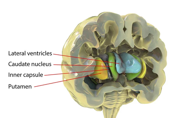
Human Brain Anatomy, Basal Ganglia. 3D Illustration Showing Caudate Nucleus (green), Putamen (yellow), And Lateral Ventricles (blue)
Image, 6.25MB, 6252 × 4168 jpg
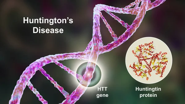
Huntington's Chorea, A Neurodegenerative Disease Due To A Mutation In The Huntingtin Gene HTT, Deficiency In The Huntingtin Protein And Changes In Brain Basal Ganglia, 3D Illustration
Image, 11.43MB, 7200 × 4050 jpg
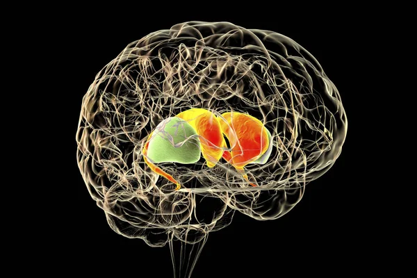
Dorsal Striatum In The Human Brain, 3D Illustration. It Is A Nucleus In The Basal Ganglia, Consists Of The Caudate Nucleus (red) And The Putamen (green), Is A Component Of The Motor And Reward Systems
Image, 6.21MB, 6000 × 4000 jpg

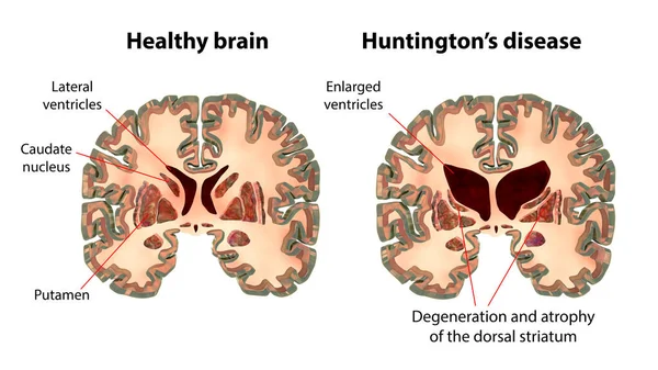
Coronal Sections Of A Healthy Brain And A Brain In Huntington's Disease Showing Enlarged Anterior Horns Of The Lateral Ventricles, Degeneration And Atrophy Of The Dorsal Striatum, 3D Illustration
Image, 10.57MB, 11104 × 6246 jpg


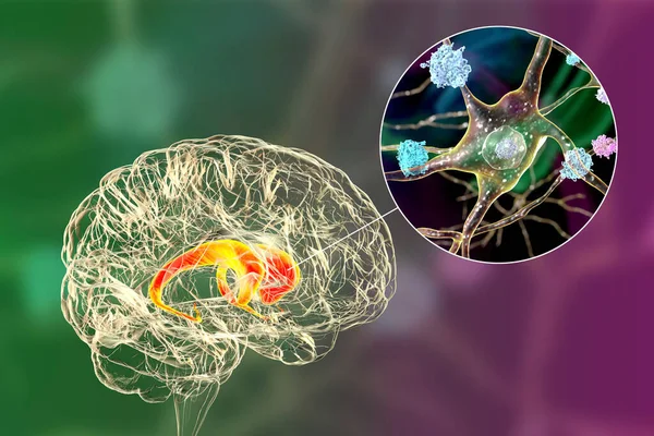
Dorsal Striatum, Caudate Nucleus And Putamen, Highlighted In The Brain Of A Person With Huntington's Disease And Close-up View Of Neuronal Degradation, Conceptual 3D Illustration
Image, 12.32MB, 7814 × 5210 jpg


Sick Little Child Girl With Epileptic Seizures In Outdoor Park,daughter Suffering From Seizures,illness With Epilepsy During Seizure Attack,asian Mother,father Care Of Girl Patient,brain,nervous System Concept
Image, 7.73MB, 5472 × 3648 jpg
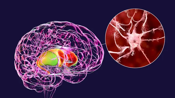
Dorsal Striatum Highlighted In Human Brain And Close-up View Of Degrading Neurons Of Dorsal Striatum Seen In Huntington's Disease, 3D Illustration
Image, 16.37MB, 8672 × 4878 jpg
Trembling Linear Icon. Anxiety. Shaking Body. Thin Line Illustration. Worrying And Afraid Person. Chills. Physiological Stress Symptom. Contour Symbol. Vector Isolated Outline Drawing. Editable Stroke
Vector, 0.56MB, 5000 × 5000 eps
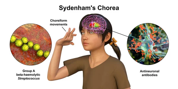
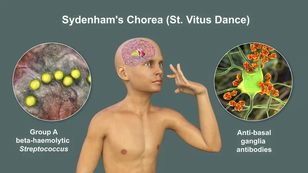
Sydenham's Chorea, An Autoimmune Disease That Results From Streptococcus Infection, Formation Of Anti-neuronal Antibodies Damaging Brain Basal Ganglia That Cause Involuntary Movements, 3D Illustration
Image, 12.54MB, 8488 × 4244 jpg
Trembling Glyph Icon. Silhouette Symbol. Anxiety. Shaking Body. Worrying And Afraid Person. Chills. Physiological Stress Symptoms. Negative Space. Vector Isolated Illustration
Vector, 0.55MB, 5000 × 5000 eps
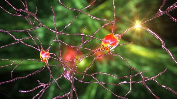
Neurons Of Dorsal Striatum, 3D Illustration. The Dorsal Striatum Is A Nucleus In The Basal Ganglia, Degrading Of Its Neurons Plays A Crucial Role In The Development Of Huntington's Disease
Image, 10.79MB, 7200 × 4050 jpg
Sydenham's Chorea, An Autoimmune Disease That Results From Streptococcus Infection, Formation Of Anti-neuronal Antibodies Damaging Brain Basal Ganglia That Cause Involuntary Movements, 3D Illustration
Image, 16.36MB, 8737 × 4914 jpg
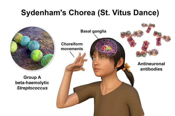
Sydenham's Chorea, An Autoimmune Disease That Results From Streptococcus Infection, Formation Of Anti-neuronal Antibodies Damaging Brain Basal Ganglia That Cause Involuntary Movements, 3D Illustration
Image, 10.04MB, 7883 × 5255 jpg
Tardive Dyskinesia Icon. Tremor From Medication. Movement Problem From Neuroleptics. Chorea, Athetosis. Mental Disorder. Flat Design, Linear And Color Styles. Isolated Vector Illustrations
Vector, 1.43MB, 5000 × 5000 eps
Neuronal Inclusions In The Caudate Nucleus Of The Brain In Huntington's Disease, 3D Illustration. Inclusions Are Composed Of Mutated Huntingtin Protein, They Are Found In Nuclei, Axons And Dendrites
Image, 13.03MB, 7996 × 5331 jpg

Dorsal Striatum Highlighted In Child's Brain And Close-up View Of Its Neurons, 3D Illustration. It Is A Nucleus In The Basal Ganglia, A Component Of The Motor And Reward Systems
Image, 31.18MB, 9339 × 6226 jpg
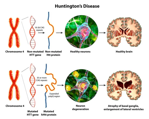
Molecular Genesis Of Huntington's Disease, 3D Illustration. Expansion Of The CAG Trinucleotide Sequence In The Htt Gene Causes Production Of Mutated Huntingtin Protein Leading To Neurodegeneration
Image, 17.01MB, 12941 × 10352 jpg

Caregiver Woman Holding Hands To Elderly With Alzheimer Disease At Home,Adult Social Care Concept
Image, 18.37MB, 8400 × 5608 jpg

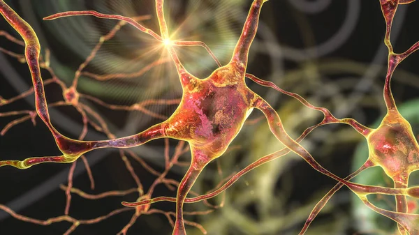
Neurons Of Dorsal Striatum, 3D Illustration. The Dorsal Striatum Is A Nucleus In The Basal Ganglia, Degrading Of Its Neurons Plays A Crucial Role In The Development Of Huntington's Disease
Image, 12.53MB, 7200 × 4050 jpg

Woman Hands Holding To Elderly With Alzheimer Disease At Nature,Adult Social Care Concept
Image, 19.15MB, 9500 × 6342 jpg
Trembling App Icon. Anxiety. Shaking Body. Worrying And Afraid Person. Chills. Physiological Stress Symptoms.
Vector, 0.75MB, 5000 × 5000 eps
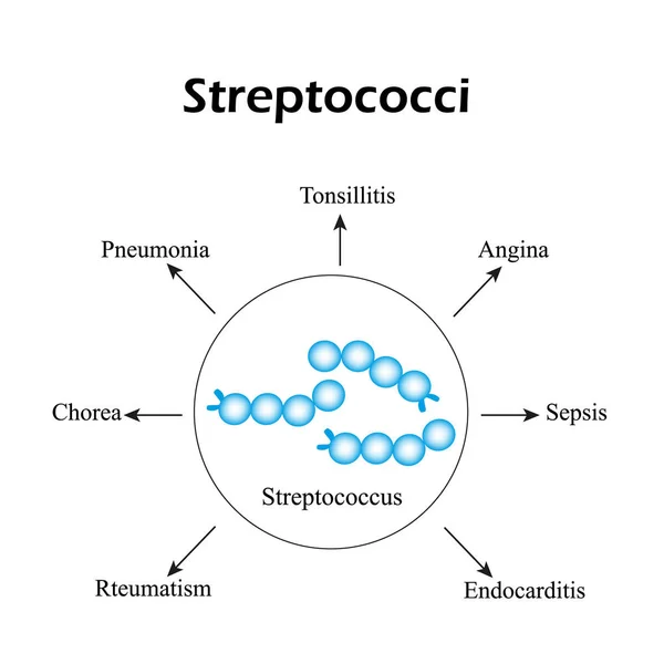
Streptococci. Streptococcal Diseases. Infographics. Vector Illustration
Vector, 3.38MB, 5000 × 5000 eps


Dorsal Striatum Highlighted In Child's Brain And Close-up View Of Its Neurons, 3D Illustration. It Is A Nucleus In The Basal Ganglia, A Component Of The Motor And Reward Systems
Image, 23.52MB, 9339 × 6226 jpg

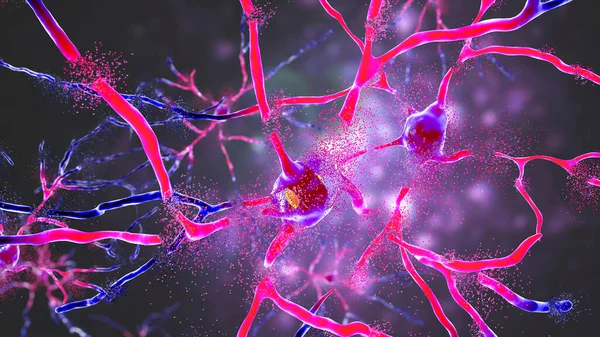
Destruction Of Neurons Of The Caudate Nucleus, Conceptual 3D Illustration. Caudate Nucleus Belongs To The Brain Basal Ganglia, Its Neurons Are Damaged In Huntingon's Disease And Other Choreas
Image, 16.49MB, 7996 × 5331 jpg

Dorsal Striatum, Caudate Nucleus And Putamen, Highlighted In The Brain Of A Person With Huntington's Disease And Close-up View Of Neuronal Inclusions, Conceptual 3D Illustration
Image, 7.98MB, 6000 × 6000 jpg

Intranuclear Neuronal Inclusions, 3D Illustration. Intranuclear Inclusions In Neurons Are Found In Different Neurodegenerative Diseases, Including Huntingon's Disease, Spinocerebellar Ataxia And Other
Image, 9.43MB, 7200 × 4050 jpg

Trembling Color Icon. Anxiety. Shaking Body. Worrying And Afraid Person. Chills. Physiological Stress Symptoms. Isolated Vector Illustration
Vector, 0.56MB, 5000 × 5000 eps
Tardive Dyskinesia Color Icon. Tremor From Medication. Movement Problem From Neuroleptics. Chorea, Athetosis. Mental Disorder. Neurological Disease From Pills. Isolated Vector Illustration
Vector, 0.63MB, 5000 × 5000 eps
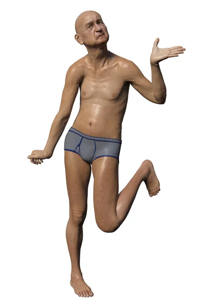
A Person With Huntington's Chorea, A Neurodegenerative Disease Due To Mutation In The HTT Gene, Changes In Brain Basal Ganglia Leading To Random Uncontrollable Movements And Dementia, 3D Illustration
Image, 3.8MB, 3297 × 4946 jpg

Neuronal Inclusions In Huntington's Disease, 3D Illustration. Inclusions Are Composed Of Mutated Huntingtin Protein, They Are Initially Formed At Axons And Dendrites, Then Migrate To Nuclei Of Neurons
Image, 9.3MB, 7200 × 4050 jpg

Anti-basal Ganglia Antibodies. 3D Conceptual Illustration Showing Molecules Of Immunoglobulins Attacking Dorsal Striatum Highlighted In The Child's Brain. They Are Found In Post-rheumatic Fever Chorea
Image, 25.08MB, 9828 × 6552 jpg
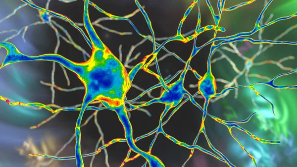
Neurons Of Dorsal Striatum, 3D Illustration. The Dorsal Striatum Is A Nucleus In The Basal Ganglia, Degrading Of Its Neurons Plays A Crucial Role In The Development Of Huntington's Disease
Image, 8.02MB, 7200 × 4050 jpg

Caudate Nuclei In Human Brain And Its Neurons, 3D Illustration. The Caudate Nucleus Is A Component Of The Basal Ganglia, It Plays Role In Choreas, Neurodegenerative And Other Brain Diseases
Image, 12.8MB, 8157 × 5438 jpg
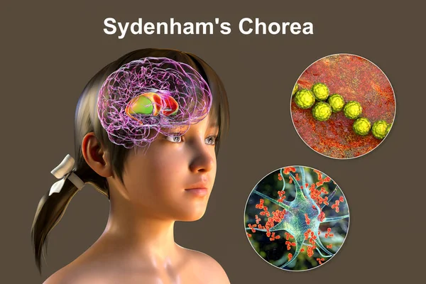
Sydenham's Chorea, An Autoimmune Disease That Results From Streptococcus Infection, Formation Of Anti-neuronal Antibodies Damaging Brain Basal Ganglia That Cause Involuntary Movements, 3D Illustration
Image, 39.11MB, 10609 × 7073 jpg
Neurons Of Dorsal Striatum, 3D Illustration. Dorsal Striatum Is A Nucleus In The Basal Ganglia, Degrading Of Its Neurons Plays Crucial Role In Development Of Huntington's Disease
Image, 12.86MB, 7200 × 4050 jpg
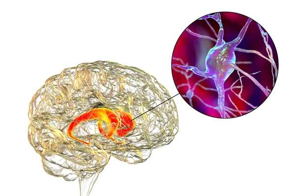
Caudate Nuclei In Human Brain And Its Neurons, 3D Illustration. The Caudate Nucleus Is A Component Of The Basal Ganglia, It Plays Role In Choreas, Neurodegenerative And Other Brain Diseases
Image, 11.51MB, 7996 × 5331 jpg

Neurons Of Dorsal Striatum, 3D Illustration. Dorsal Striatum Is A Nucleus In The Basal Ganglia, Degrading Of Its Neurons Plays Crucial Role In Development Of Huntington's Disease
Image, 8.32MB, 7200 × 4050 jpg

Brain Dorsal Striatum Anatomy, 3D Illustration. The Dorsal Striatum Consists Of The Caudate Nucleus (orange) And The Putamen (blue). Amygdala Is Colored In Red. Front View
Image, 5.28MB, 6000 × 4000 jpg

Intranuclear Neuronal Inclusions, 3D Illustration. Intranuclear Inclusions In Neurons Are Found In Different Neurodegenerative Diseases, Including Huntingon's Disease, Spinocerebellar Ataxia And Other
Image, 8.23MB, 7200 × 4050 jpg

Sydenham's Chorea, An Autoimmune Disease That Results From Streptococcus Infection, Formation Of Anti-neuronal Antibodies Damaging Brain Basal Ganglia That Cause Involuntary Movements, 3D Illustration
Image, 10.26MB, 6947 × 4632 jpg

A Boy With Sydenham's Chorea And Involuntary Movements Of A Hand, 3D Illustration. An Autoimmune Disease After Streptococcus Infection Due To Antibodies Against Cells Of Brain Basal Ganglia And Heart
Image, 8.84MB, 6031 × 4021 jpg
Page 1 >> Next