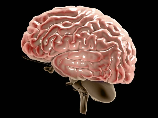
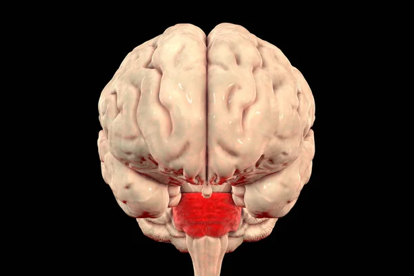
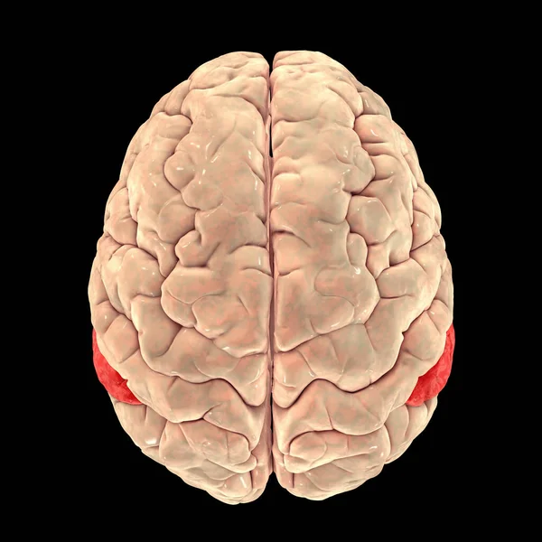
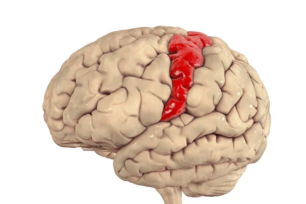
Stock image Human brain nervous system anatomy, sections are separated by colored spots, medical diagram with parasympathetic and sympathetic nerves. medically accurate, Central organ, 3d render
Published: Nov.28, 2022 10:40:52
Author: aamine29000
Views: 3
Downloads: 0
File type: image / jpg
File size: 8.68 MB
Orginal size: 7979 x 4488 px
Available sizes:
Level: beginner