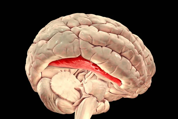
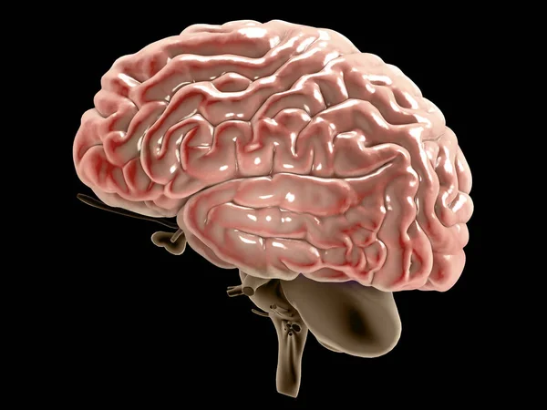
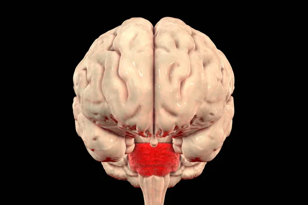
Stock image Pons Varolii highlighted inside human brain, 3D illustration. It part of the brainstem, includes neural pathways carrying signals from the brain down to cerebellum and medulla, and into the thalamus
Published: Dec.03, 2020 14:34:00
Author: katerynakon
Views: 17
Downloads: 0
File type: image / jpg
File size: 3.75 MB
Orginal size: 6000 x 4000 px
Available sizes:
Level: silver