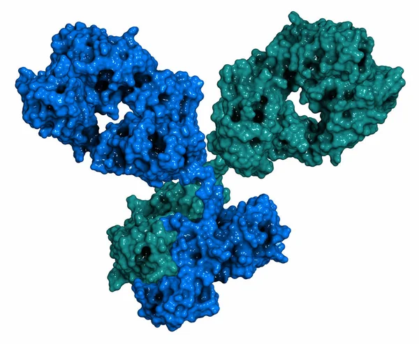
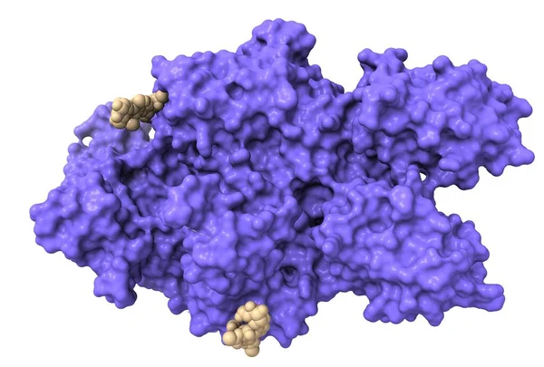
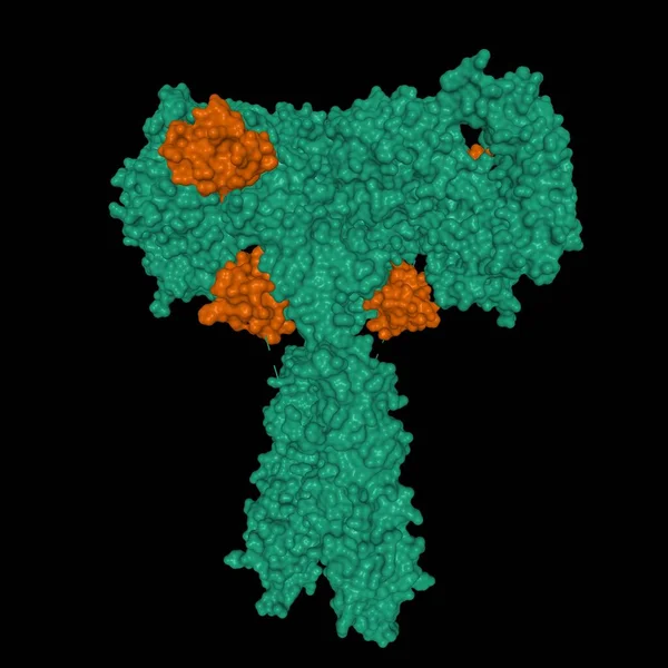
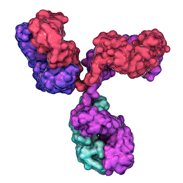
Stock image IgG2a monoclonal antibody (immunoglobulin). 3D Illustration. Many biotech drugs are antibodies.

Published: Aug.13, 2021 16:10:03
Author: Wirestock
Views: 23
Downloads: 1
File type: image / jpg
File size: 11.66 MB
Orginal size: 8000 x 6000 px
Available sizes:
Level: platinum