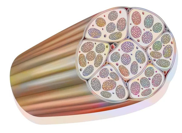
Stock image Illustration of the anatomy of a typical peripheral nerve.

Published: Jun.10, 2021 06:53:43
Author: aldonagriskeviciene
Views: 3
Downloads: 0
File type: image / jpg
File size: 3.82 MB
Orginal size: 5728 x 5302 px
Available sizes:
Level: beginner