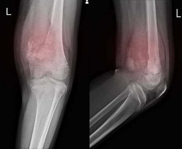
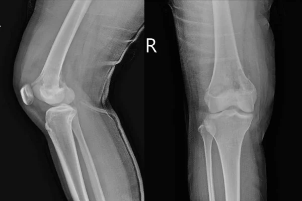
Stock image Knee joint.No fracture, dislocation , bony destruction .
Published: Aug.20, 2019 14:03:58
Author: Richmanphoto
Views: 221
Downloads: 2
File type: image / jpg
File size: 12.44 MB
Orginal size: 7862 x 4986 px
Available sizes:
Level: bronze