Home
Images
Vectors
Videos
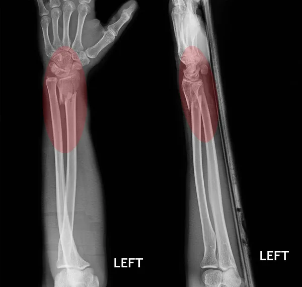
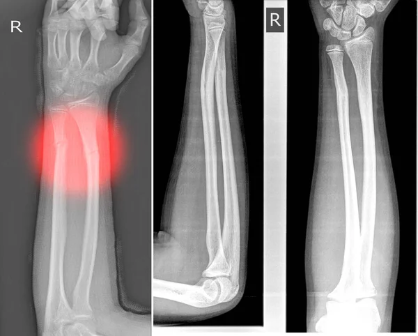
Stock image X-ray image right of wrist joint, shows step fracture of the distal radius and ulna
Published:
Mar.03, 2020 11:39:37
Author:
Richmanphoto
Views:
13
Downloads:
0
File type:
image
/
jpg
File size:
2.96 MB
Orginal size:
3664
x
2950
px
Available sizes:
S
(500 x 403)
M
(1000 x 805)
L
(2000 x 1610)
XL
(3664 x 2950)
EL0
(3664 x 2950)
Level:
bronze
Download Image
Image topics:
Image
Background
Human
People
Health
Medicine
Healthcare
Medical
Care
Technology
Hand
Wrist
Clinical
Doctor
Hospital
Fragile
Bone
Finger
Exam
Patient
Treatment
Broken
Film
Pain
Joint
Skeleton
Fracture
Diagnosis
Anatomy
Radius
X Ray
Surgery
Trauma
Accident
Injury
Scan
Operation
Radiology
Painful
Forearm
Deformity
Radiograph
Orthopedic
Carpal
Extremity
Ulna
Distal
Wrist Fracture
Distal End Radius
Distal End Radius Fracture
Similar stock images
Fracture Shaft Of Radius & Ulnar Bone, X-ray Film
4206 × 4000
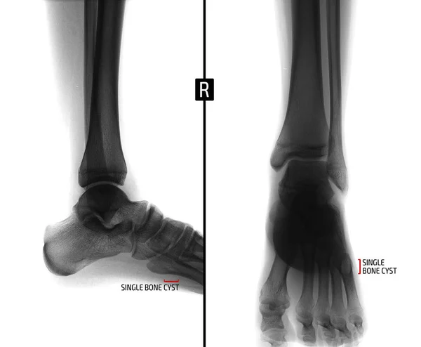
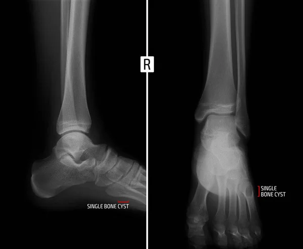
X-ray Of The Ankle Joint. Shows The Bony Cyst Of The Fifth Finger Of The Right Foot. Marker. Negative.
2976 × 2454
X-ray Foot And Ankle Normal Joint And Osteochondroma Of Distal Tibia Cause Pressure Effect To Fibula.Medical Image Concept.
6000 × 4000
X-ray Of The Ankle Joint. Shows The Bony Cyst Of The Fifth Finger Of The Right Foot. Marker.
2976 × 2454
X-ray Foot And Ankle (AP,Lat)showing Metatarsal Fractures On Red Point.Metatarsal Fractures Are Among The Most Common Injuries Of The Foot.
4000 × 2950
Film X-ray Wrist Fracture : Show Fracture Radius Bone (forearm's
4903 × 3282
Fracture Shaft Of Radius & Ulnar Bone, X-ray Film
4206 × 4000
Double Fracture Of Tibia, Two Projections, X-ray
3616 × 2682