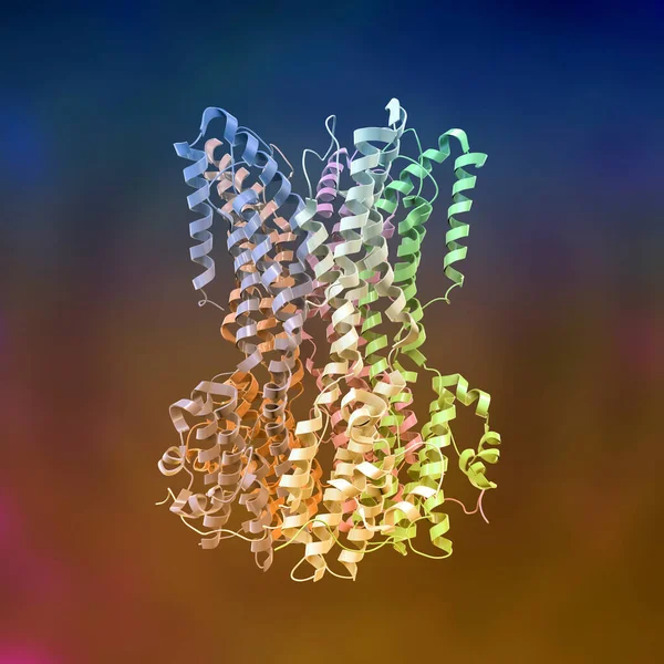
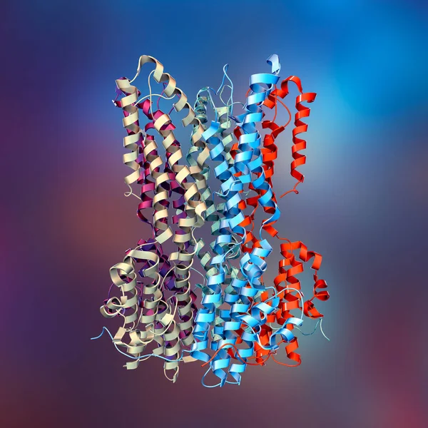
Stock image Molecular model of Bestrophin-1 (Best 1) protein, 3D illustration. A protein responsible for regulating calcium signaling in cells. Mutation of Best1 causes a group of degenerative retinal diseases
Published: Mar.16, 2023 08:26:23
Author: katerynakon
Views: 5
Downloads: 0
File type: image / jpg
File size: 12.68 MB
Orginal size: 6624 x 6624 px
Available sizes:
Level: silver