
Stock image Bestrophin
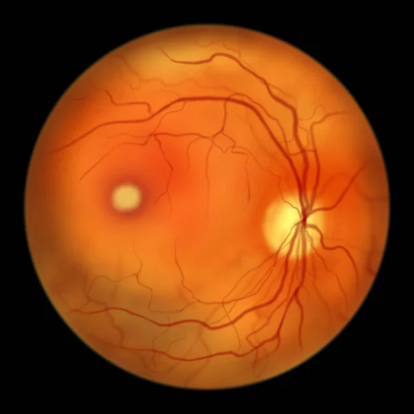
Best Disease. Best Vitelliform Macular Dystrophy, Vitelliform Stage, Classic Egg-yolk Lesion, Scientific Illustration, Ophthalmoscope View
Image, 2.87MB, 5000 × 5000 jpg
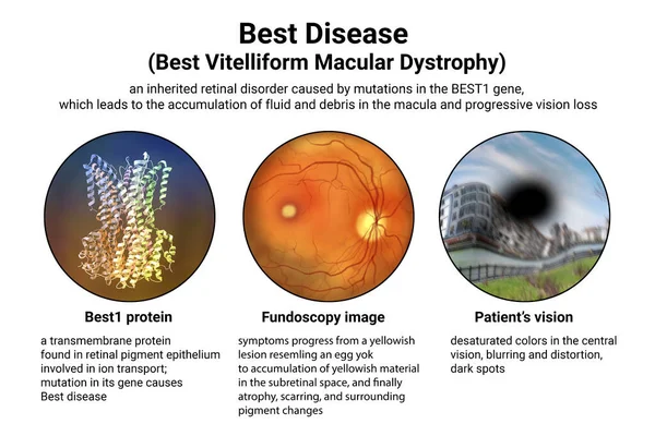
Best Disease. Best Vitelliform Macular Dystrophy, 3D Illustration Showing Best1 Protein, Classic Fundoscopic Egg-yolk Lesion On Retina, And Distorted Vision With Black Spot In A Patient
Image, 7.56MB, 9000 × 6000 jpg

Best Disease. Best Vitelliform Macular Dystrophy, Pseudohypopyon Stage, Layering Of Lipofuscin, Scientific Illustration, Ophthalmoscope View
Image, 2.89MB, 5000 × 5000 jpg

Best Disease, Illustration Showing Normal Eye Retina And Best Vitelliform Macular Dystrophy, Vitelleruptive Stage With Scrambled Egg Appearance On Fluorescein Angiography
Image, 8.14MB, 11738 × 6603 jpg
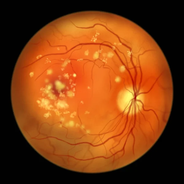
Autosomal Recessive Bestrophinopathy, Ophthalmoscope View, Scientific Illustration Showing Accumulation Of Lipofuscin Deposits Around And Beyond The Macula Leading To Progressive Damage To The Retina
Image, 3MB, 5000 × 5000 jpg

The Difference Between The Vision Of A Normal Eye And An Eye Affected By Best Disease, Illustration Showing Desatured Colors In The Central Vision, Blurring, Distortion, Black Spot
Image, 12.33MB, 9000 × 6000 jpg
Page 1 >> Next