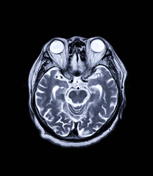
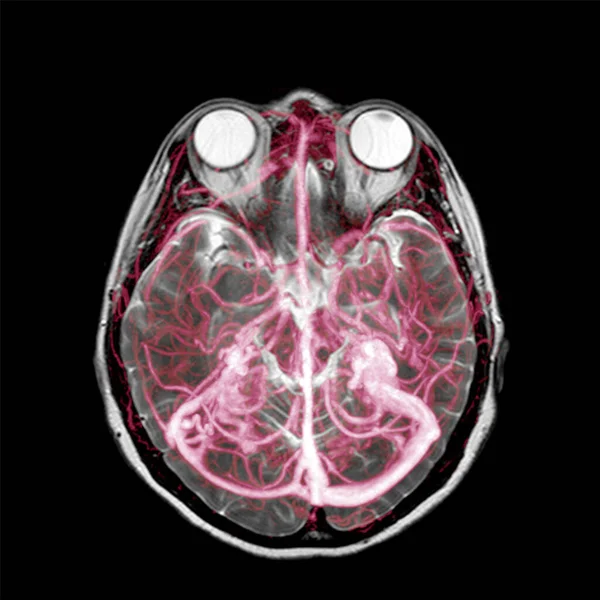
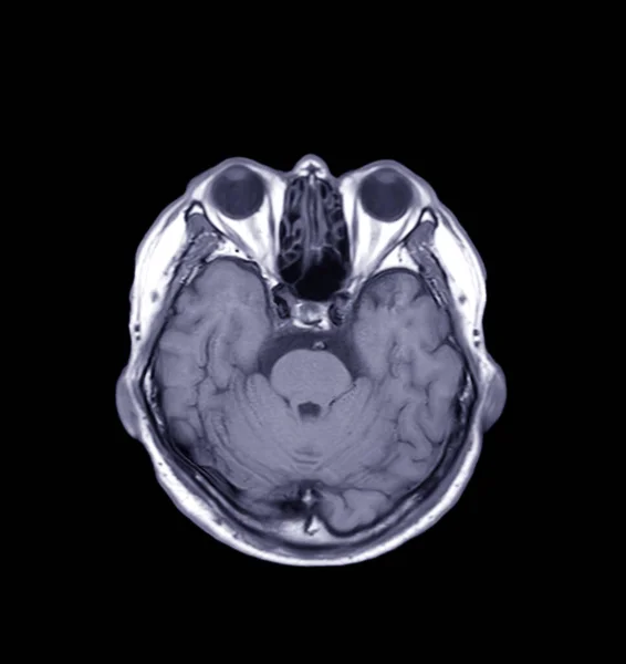
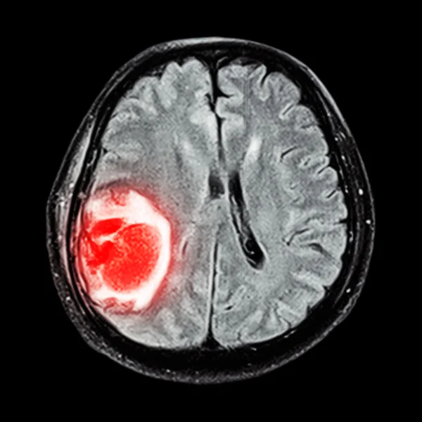
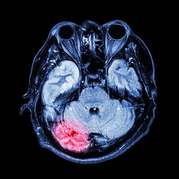
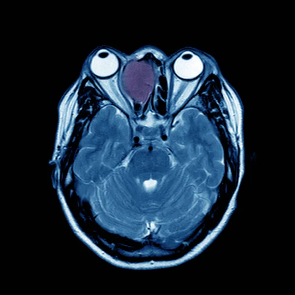
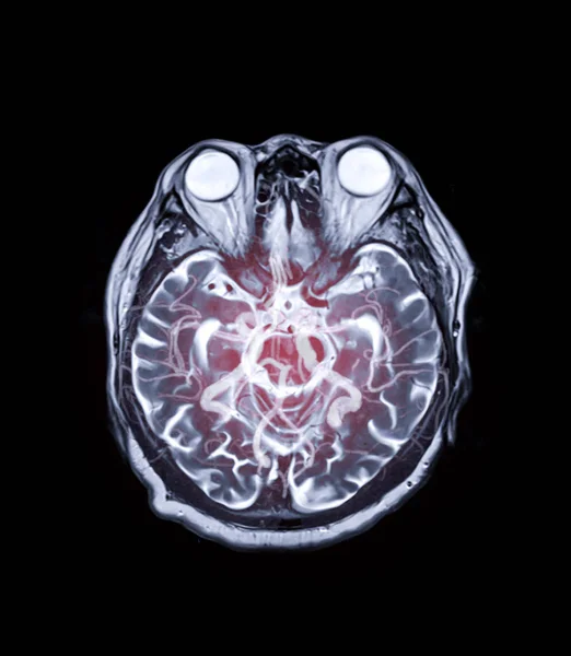
Stock image MRI brain Axial T2 technique with mra brain mix image for background concept.
Published: Dec.23, 2019 11:43:28
Author: samunella
Views: 16
Downloads: 0
File type: image / jpg
File size: 1.91 MB
Orginal size: 2904 x 3339 px
Available sizes:
Level: beginner