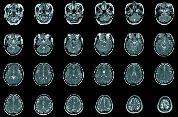
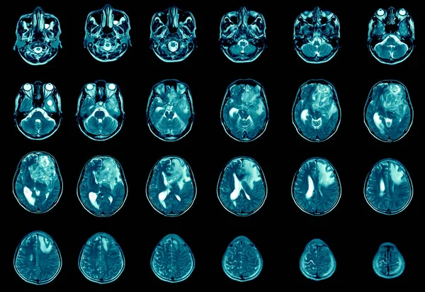
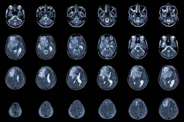
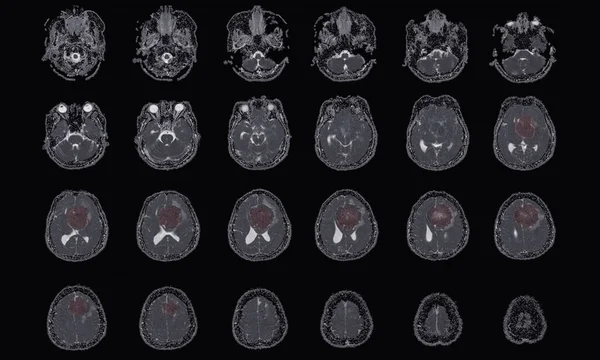
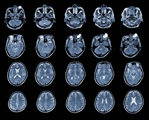
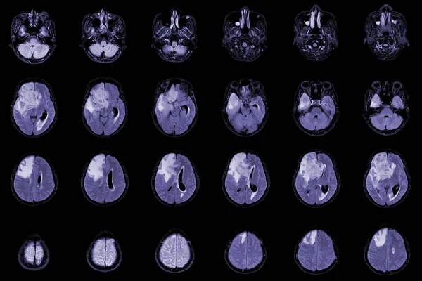
Stock image MRI Brain Axial views .to evaluate brain tumor. Glioblastoma, brain metastasis isodensity mass with an ill-defined margin and surrounding edema at the right frontal lobe.
Published: Dec.05, 2022 08:02:24
Author: Richmanphoto
Views: 6
Downloads: 0
File type: image / jpg
File size: 4.76 MB
Orginal size: 6000 x 4000 px
Available sizes:
Level: bronze