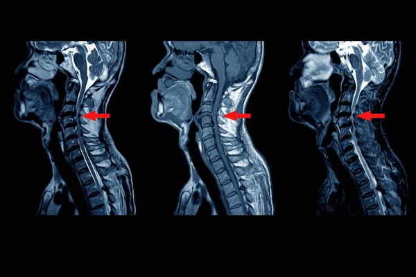
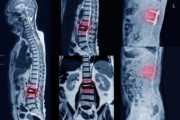
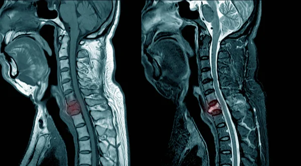
Stock image MRI of cervical spine compression fracture at C7.
Published: Jul.15, 2019 12:03:32
Author: Richmanphoto
Views: 555
Downloads: 4
File type: image / jpg
File size: 4.6 MB
Orginal size: 6280 x 3458 px
Available sizes:
Level: bronze