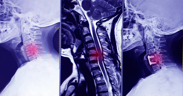
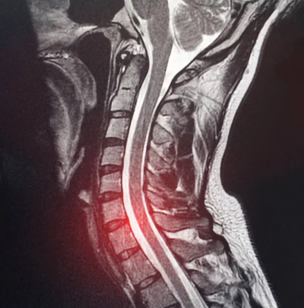
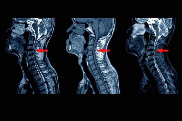
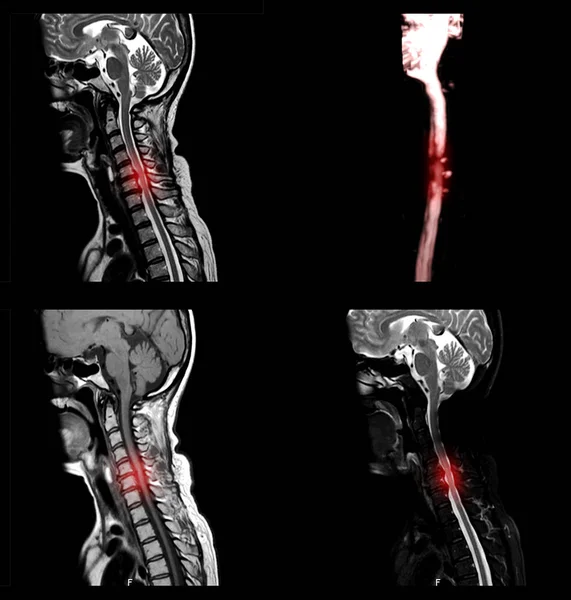
Stock image MRI OF CERVICAL SPINE : Moderate to severe posterior central disc protrusion of C3/4 to C5/6 intervertebral discs with a 2.0 cm in length small posterior subligamentous fluid collection.on red point
Published: Jun.03, 2020 09:13:40
Author: Richmanphoto
Views: 65
Downloads: 5
File type: image / jpg
File size: 4.36 MB
Orginal size: 6000 x 4000 px
Available sizes:
Level: bronze
Similar stock images


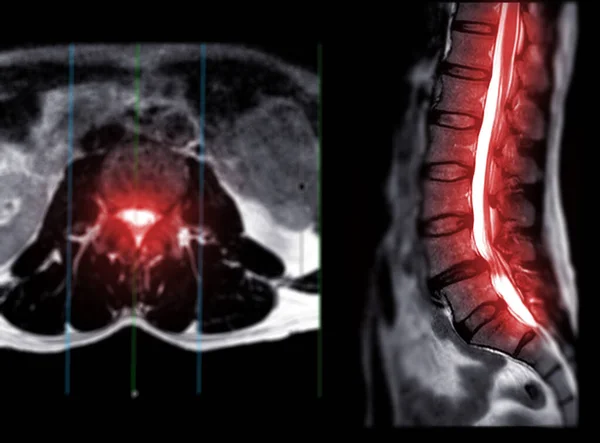
MRI L-S Spine Or Lumbar Spine Axial T2W View With Sagittal Plane For Diagnosis Spinal Cord Compression.
3032 × 2240