Home
Images
Vectors
Videos
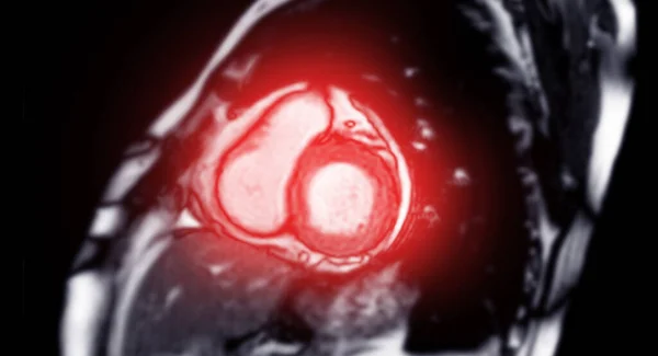
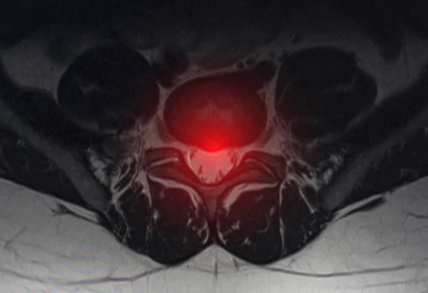
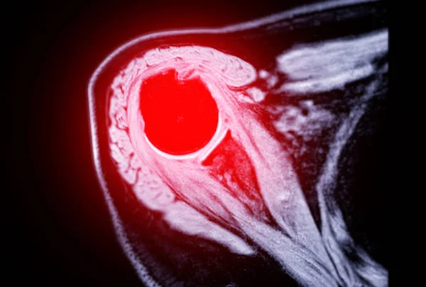
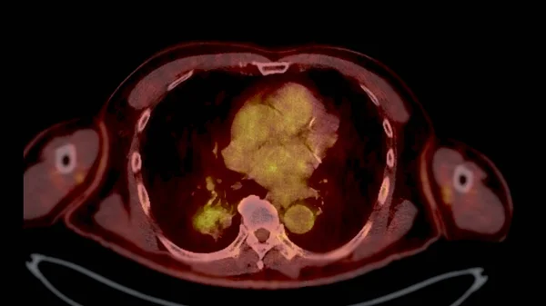
Stock image MRI of the prostate gland reveals a focal abnormal signal intensity (SI) lesion at the left posterolateral peripheral zones at the apex, aiding in diagnosing tumors and guiding treatment decisions.
Published:
Apr.27, 2024 04:06:32
Author:
samunella
Views:
0
Downloads:
0
File type:
image
/
jpg
File size:
3.55 MB
Orginal size:
4056
x
3205
px
Available sizes:
S
(500 x 395)
M
(1000 x 790)
L
(2000 x 1580)
XL
(4056 x 3205)
EL0
(4056 x 3205)
Level:
beginner
Download Image
Image topics:
Human
Health
Medicine
Illness
Male
Medical
Hospital
Patient
Treatment
Aged
Disease
Science
Cancer
Magnetic
Diagnosis
Examination
Anatomy
Imaging
Resonance
Scanner
Disorder
Awareness
Prognosis
Mens
Tumor
Apex
Reproductive
Bladder
Localization
Biopsy
Urethra
Glandular
MRI Scan
Health Care
Anatomy Body
Prostatitis
Prostate Cancer
Sagittal
Prostate Gland
Prostatic
Urinary System
Early Detection
Enlarged Prostate
Management Planning
Mri Prostate
Mri Lower Abdomen
Left Posterolateral Peripheral Zones
Signal Intensity Lesion
Treatment Decisions
Focal Abnormality
Similar stock images
MRI Heart Or Cardiac MRI ( Magnetic Resonance Imaging ) Of Heart Short Axis For Diagnosis Heart Disease.
6060 × 3284
Image After Examination Of Magnetic Resonance Imaging. Spine Examination Concept, Degenerative Changes In The Intervertebral Discs, Spondylarthrosis
4510 × 3093
Magnetic Resonance Imaging Or MRI Of Shoulder Joint For Diagnostic Shoulder Pain.
6144 × 4152
PET CT Scan Fusion Image It Provides Detailed Images By Merging Metabolic Activity From PET With Anatomical Information From CT Scans.
3840 × 2160
PET CT Scan Fusion Image It Provides Detailed Images By Merging Metabolic Activity From PET With Anatomical Information From CT Scans.
3840 × 2160
MRI Heart Or Cardiac MRI ( Magnetic Resonance Imaging ) Of Heart Short Axis View For Diagnosis Heart Disease.
4320 × 3604
Fantasy Chaotic Colorful Fractal Pattern. Abstract Fractal Shapes. 3D Rendering Illustration Background Or Wallpaper.
3840 × 2160
Magnetic Resonance Imaging Or MRI Of Knee Joint Axial T2 FS View For Detect Tear Or Sprain Of The Anterior Cruciate Ligament (ACL)
5190 × 3731