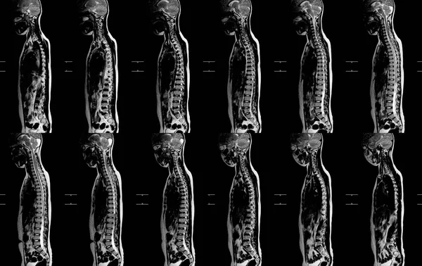
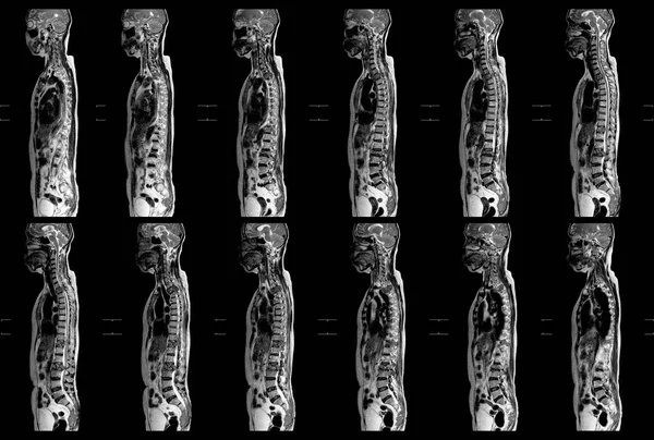
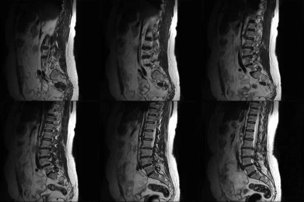
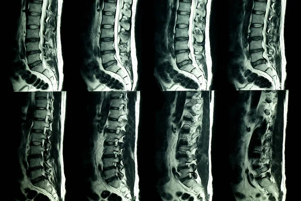
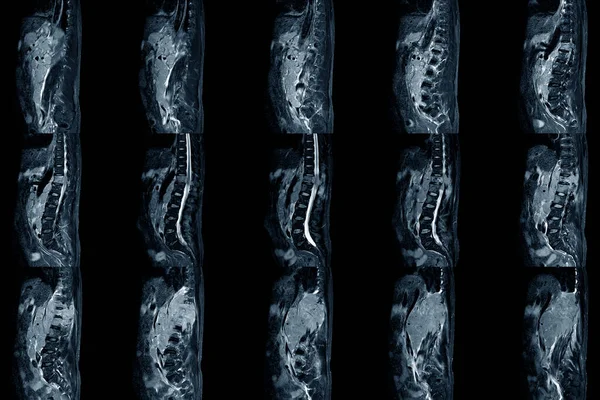
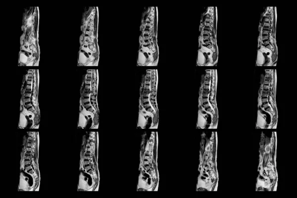
Stock image MRI OF THE THORACOLUMBAR SPINE a patient who has moderate to severe compression fractures of T11 and L2 levels with abnormal signal intensity view of science and education.
Published: Dec.05, 2022 08:02:24
Author: Richmanphoto
Views: 2
Downloads: 0
File type: image / jpg
File size: 4.51 MB
Orginal size: 6000 x 4000 px
Available sizes:
Level: bronze