Home
Images
Vectors
Videos
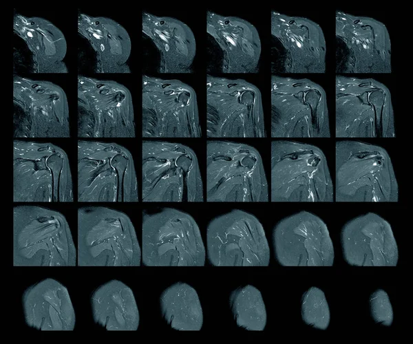
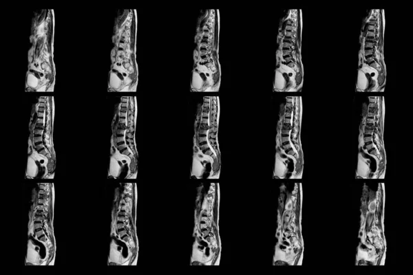
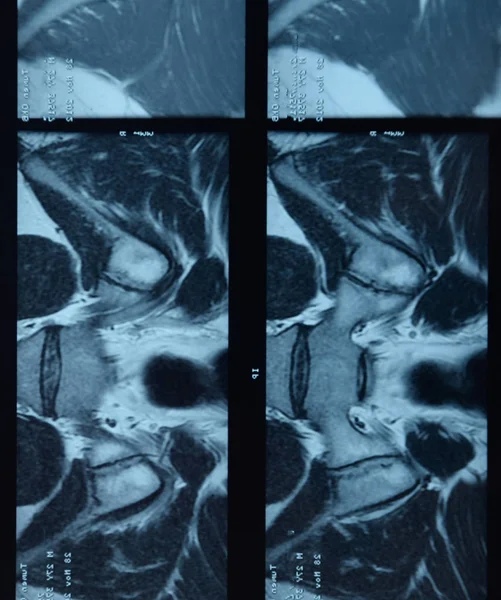
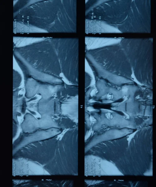
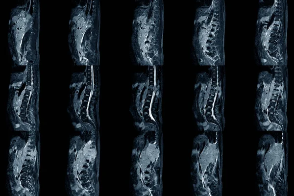
Stock image Lumbar spinal stenosis MRI scan Sagittal view finding moderate posterior inferior tumor protrusion cause bilateral root compression. Chronic low back pain disease
Published:
Oct.27, 2021 08:54:29
Author:
Richmanphoto
Views:
3
Downloads:
0
File type:
image
/
jpg
File size:
7.79 MB
Orginal size:
6000
x
4000
px
Available sizes:
S
(500 x 333)
M
(1000 x 667)
L
(2000 x 1333)
XL
(6000 x 4000)
EL0
(6000 x 4000)
Level:
bronze
Download Image
Image topics:
Human
Health
Medicine
Illness
Medical
Cord
Technology
Hospital
Bone
Exam
Patient
Body
Disease
Film
Pain
Compression
Disc
Muscle
Spine
Joint
Magnetic
Skeleton
Diagnosis
Examination
Anatomy
Spinal
Vertebra
Imaging
Surgery
Injury
Scan
Diagnostic
Resonance
Radiology
Xray
Orthopedic
Mri
Nerve
Thoracic
Degenerative
Cervical
Lumbar
Tomography
Health Care And Medicine
Medical Research
Stenosis
Cervical Spine
Magnetic Resonance Imaging
Diagnostic Imaging
Disc Disease
Similar stock images
Magnetic Resonance Imaging Of Left Shoulder Rotator Cuff Tear With Suspected Lipoma Of Left Shoulder Science And Education Mri Shoulder Background,Medical.
7732 × 6448
MRI C-Spine,neck, Image 6view Show Cervical Neck Dislocation.
6000 × 4000
MRI OF THE THORACOLUMBAR SPINE A Patient Who Has Moderate To Severe Compression Fractures Of T11 And L2 Levels With Abnormal Signal Intensity View Of Science And Education.
6000 × 4000
MRI Sacroiliac Articulation. Study Of Ankylosing Spondyloarthritis Patient.
2340 × 2800
MRI Sacroiliac Articulation. Study Of Ankylosing Spondyloarthritis Patient.
5896 × 3931
Radiograph Of The Spine
2170 × 3330
MRI Sacroiliac Articulation. Study Of Ankylosing Spondyloarthritis Patient.
2340 × 2800
X-ray Of The Lower Parts Of The Gastrointestinal Tract (GI). Tight Filling. Emptying. Insufflation By Air. Dolichocolon.
10650 × 3260